microPublication

Get Your Data Out, Be Cited

  • About
    • Editorial Policies
      • Editorial Staff
      • Editorial Board
      • Criteria For Publication
      • Publishing Information
      • Data Sharing Policy
    • For Authors
      • Preparation And Submission Of A Manuscript
      • Peer Review Process
      • Following Acceptance
      • Appeals
    • For Reviewers
    • Why micropublish?
  • Submit a microPublication
  • Journals
    • microPublication Biology
      • Editorial Board
  • microPublications
    • Biology
      • Species
        • Arabidopsis
        • C. elegans
        • D. discoideum
        • Drosophila
        • Human
        • Mouse
        • S. cerevisiae
        • S. pombe
        • Xenopus
        • Zebrafish
      • Categories
        • Phenotype Data
        • Methods
        • Expression Data
        • Genotype Data
        • Integrations
        • Genetic Screens
        • Models of Human Disease
        • Software
        • Interaction data
        • Database Updates
        • Electrophysiology Data
        • Phylogenetic Data
        • Science and Society
        • Biochemistry
  • Contact
  • More
    • Archives
    • FAQs
    • Newsletter
microPublication / Biology / LOC496300 is expressed in the...
LOC496300 is expressed in the endoderm of developing Xenopus laevis embryos
Maria E Stewart1, Kelsey M Donahue1, Elizabeth G Wilke1 and Emily T Shifley1
1Northern Kentucky University; Highland Heights, KY
Correspondence to: Emily T Shifley (shifleye1@nku.edu)
Figure 1. (A) LOC496300 is expressed in the endoderm of Xenopus laevis from NF stage 10 through 37. It becomes restricted to the midgut and hindgut during tailbud stages (~NF stg 30) and is expressed in the intestine and gall bladder of NF stage 42-43 gut tubes at differing levels (i.e. gut tube a and b). Embryos hybridized with the sense probe for LOC496300 show some non-specific staining in the head at tailbud stages, but overall no specific staining in the endoderm. NF stage 43 organ buds are marked as follows: lv, liver; p, pancreas; s, stomach; gb, gall bladder; i, intestine. (B) NF stage 10, 12, 28 and 35 embryos were sectioned to show the punctate expression pattern of LOC496300 throughout the early endoderm and in the dorsal and ventral tailbud endoderm. The numbered lines mark the approximate level of each section of the tailbud embryos and the dotted red line outlines the endoderm.

Description

Xenopus laevis is an excellent model organism for studies on vertebrate endoderm development (Womble et al., 2016; Zorn, 2009). The endoderm contributes to a number of important organs including the lungs, liver, gall bladder, pancreas, stomach and intestine. Each of these organs differentiate from the foregut, midgut, and hindgut domains of the early endoderm (Chalmers and Slack, 2000). It has recently been shown that as early as gastrulation differential fates are being specified within the endoderm (Costa et al., 2003; Rankin et al., 2018). Several studies have worked to identify markers of different fates in the developing endoderm in order to assist with research on endoderm specification, differentiation, and organogenesis, but not all of the identified genes have known functions and some are currently unclassified (Chen et al., 2003; Costa et al., 2003; Park et al., 2007; Zorn and Mason 2001).

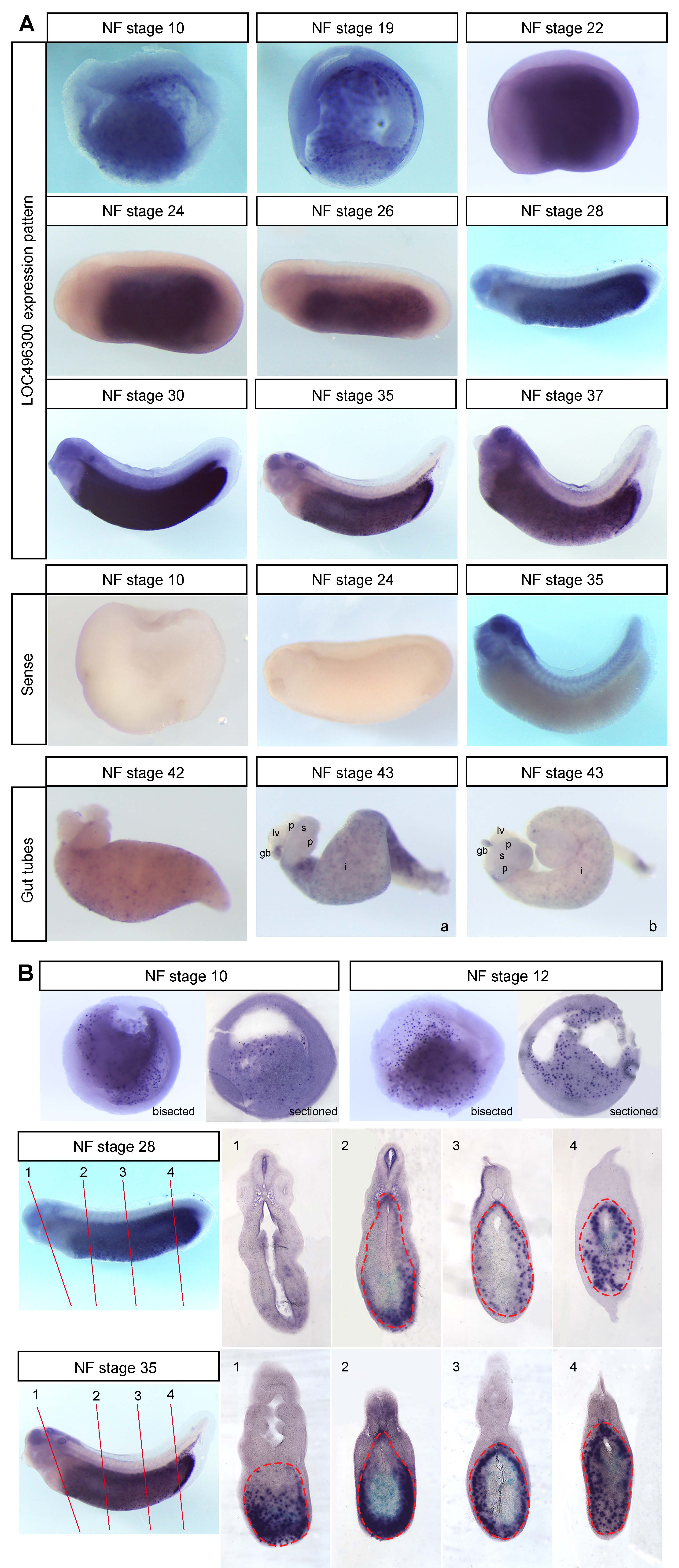
We performed whole mount in situ hybridization (WISH) on hypothetical locus LOC496300 (Xl.8755; Ref Seq NM_001095458.1) and found that it is expressed throughout the endoderm of early Xenopus embryos (NF stage 10-23) and as development proceeds it becomes localized to the midgut and hindgut endoderm (NF stage 27-37) (Figure 1A), which later gives rise to the intestine posterior to the stomach (Chalmers and Slack, 1998). LOC496300 has a similar expression pattern to other midgut-hindgut specific markers like ctbs, impdh1, and darmin during the early tadpole stages (Costa et al., 2003; Pera et al., 2003). LOC496300 does show some expression in the foregut early on, whereas darmin is excluded from the foregut even at gastrulation (Costa et al., 2003, Pera et al., 2003). We repeated the WISH for LOC496300 with three sets of embryos and saw consistent expression patterns with each set (examining a total of at least 18 embryos for each stage). WISH with the sense probe for LOC496300 as a control showed no staining in the endoderm at any stage, but non-specific staining in the brain, eye, and pharynx at NF stage 35 (see Figure 1A).

Sectioned embryos show expression of LOC496300 in the endoderm is punctate in a subset of cells throughout all regions of the early endoderm (Figure 1B). At tailbud stages LOC496300 is expressed in both the dorsal and ventral midgut and hindgut and in the gut tube at NF stage 42 and 43, maintaining a punctate expression pattern throughout the intestine. It is not clear what subset of cells might be expressing LOC496300. There are several other genes that show punctate expression patterns in the endoderm such as Sox17α, Sox17β, and darmin at NF stage 10 when there are many yolk granules still present in the endoderm cells (Costa et al., 2003; Hudson et al., 1997; Sinner et al., 2004). Later in development, it is possible LOC496300 is expressed in a group of cells that will differentiate into a specific cell type for the functional intestinal epithelium of the feeding tadpole, which consists mainly of principal absorptive cells as well as some gland cells, endocrine cells and lymphocytes (Marshall and Dixon; 1978; Shi and Ishizuya-Oka, 1996). This larval epithelium will eventually undergo apoptosis except for a few cells that de-differentiate into progenitor stem cells which give rise to the adult intestinal epithelium at metamorphosis (Ishizuya-Oka, 1996; Ishizuya-Oka, 2011; Ishizuya-Oka et al., 2009; Shi and Ishizuya-Oka, 1996; Sun et al., 2013). It would be interesting to determine which genes expressed in the embryonic intestine, like LOC496300, play a role in larval intestinal development and function, and for the dramatic transition that occurs during metamorphosis.

LOC496300 is currently unclassified. Searching for conserved domains identified a Spc7 kinetochore domain in LOC496300, suggesting it may play a role in kinetochore function. It also has KOG2044 and SbcC exonuclease domains, suggesting a possible role in replication, recombination, or repair. Further research into the functions of LOC496300 will help identify the role it plays specifically in the endoderm during development and whether it contributes to the specification and differentiation of the intestinal fate.  Additionally, it would be interesting to discover whether LOC496300 is regulated by dynamic Wnt, BMP, and RA signals during different stages of development, as others have shown that coordination of these signaling pathways specifies different endodermal fates (Rankin et al., 2018; Stevens et al., 2017).

Reagents

Xenopus laevis embryos were cultured and gut tubes were isolated using standard protocols approved by the NKU Institutional Animal Care and Use Committee (IACUC Protocol #2014-09). The plasmid to make the WISH probe for LOC496300 was purchased sequence-verified from Source Bioscience (CF286219 clone #IRBHp990A1273D) and we used SalI to linearize the plasmid and T7 to transcribe the anti-sense RNA probe (NotI with SP6 for the sense probe). We followed previously described protocols for WISH (Costa et al., 2003). For sectioning, the embryos were embedded in gelatin-albumin and cut into 50uM sections using a Leica vibratome VT1200 S.

Acknowledgments

Thank you to all the members of the Shifley lab for their assistance.

References

Chalmers AD, Slack JM. 1998. Development of the gut in Xenopus laevis. Developmental dynamics : an official publication of the American Association of Anatomists 212: 509-21.
PubMed
Chalmers AD, Slack JM. 2000. The Xenopus tadpole gut: fate maps and morphogenetic movements. Development 127: 381-92.
PubMed
Chen Y, Jurgens K, Hollemann T, Claussen M, Ramadori G, Pieler T. 2003. Cell-autonomous and signal-dependent expression of liver and intestine marker genes in pluripotent precursor cells from Xenopus embryos. Mechanisms of development 120: 277-88.
PubMed
Costa RM, Mason J, Lee M, Amaya E, Zorn AM. 2003. Novel gene expression domains reveal early patterning of the Xenopus endoderm. Gene Expr Patterns 3: 509-19.
PubMed
Hudson C, Clements D, Friday RV, Stott D, Woodland HR. 1997. Xsox17alpha and -beta mediate endoderm formation in Xenopus. Cell 91: 397-405.
PubMed
Ishizuya-Oka A. 1996. Apoptosis of larval cells during amphibian metamorphosis. Microsc Res Tech 34: 228-35.
PubMed
Ishizuya-Oka A. 2011. Amphibian organ remodeling during metamorphosis: insight into thyroid hormone-induced apoptosis. Dev Growth Differ 53: 202-12.
PubMed
Ishizuya-Oka A, Hasebe T, Buchholz DR, Kajita M, Fu L, Shi YB. 2009. Origin of the adult intestinal stem cells induced by thyroid hormone in Xenopus laevis. FASEB J 23: 2568-75.
PubMed | PubMed Central
Marshall JA, Dixon KE. 1978. Cell specialization in the epithelium of the small intestine of feeding Xenopus laevis tadpoles. Journal of anatomy 126: 133-44.
PubMed | PubMed Central
Park EC, Hayata T, Cho KW, Han JK. 2007. Xenopus cDNA microarray identification of genes with endodermal organ expression. Developmental dynamics : an official publication of the American Association of Anatomists 236: 1633-49.
PubMed
Pera EM, Martinez SL, Flanagan JJ, Brechner M, Wessely O, De Robertis EM. 2003. Darmin is a novel secreted protein expressed during endoderm development in Xenopus. Gene Expr Patterns 3: 147-52.
PubMed
Rankin SA, McCracken KW, Luedeke DM, Han L, Wells JM, et al. 2018. Timing is everything: Reiterative Wnt, BMP and RA signaling regulate developmental competence during endoderm organogenesis. Developmental biology 434: 121-32.
PubMed | PubMed Central
Shi YB, Ishizuya-Oka A. 1996. Biphasic intestinal development in amphibians: embryogenesis and remodeling during metamorphosis. Curr Top Dev Biol 32: 205-35.
PubMed
Sinner D, Rankin S, Lee M, Zorn AM. 2004. Sox17 and beta-catenin cooperate to regulate the transcription of endodermal genes. Development 131: 3069-80.
PubMed
Stevens ML, Chaturvedi P, Rankin SA, Macdonald M, Jagannathan S, et al. 2017. Genomic integration of Wnt/beta-catenin and BMP/Smad1 signaling coordinates foregut and hindgut transcriptional programs. Development 144: 1283-95.
PubMed | PubMed Central
Sun G, Heimeier RA, Fu L, Hasebe T, Das B, et al. 2013. Expression profiling of intestinal tissues implicates tissue-specific genes and pathways essential for thyroid hormone-induced adult stem cell development. Endocrinology 154: 4396-407.
PubMed | PubMed Central
Womble M, Pickett M, Nascone-Yoder N. 2016. Frogs as integrative models for understanding digestive organ development and evolution. Seminars in cell & developmental biology 51: 92-105.
PubMed | PubMed Central
Zorn A. 2009. Endoderm Development and Organ Formation. Annu Rev Cell Dev Biol.
PubMed
Zorn AM, Mason J. 2001. Gene expression in the embryonic Xenopus liver. Mechanisms of development 103: 153-7.
PubMed

Funding

This work was supported by the Kentucky Biomedical Research Infrastructure Network NIGMS grant #8P20GM103436 as well as the Northern Kentucky University Research Foundation, Northern Kentucky University Faculty Senate, and NKU Center for Integrative Natural Science and Mathematics (CINSAM).

Author Contributions

Maria E Stewart: Investigation, Visualization
Kelsey M Donahue: Investigation, Visualization
Elizabeth G Wilke: Investigation
Emily T Shifley: Conceptualization, Writing - original draft, Writing - review and editing, Supervision.

Reviewed By

Sang-Wook Cha and Christina James-Zorn

History

Received: June 26, 2019
Accepted: August 2, 2019
Published: August 12, 2019

Copyright

© 2019 by the authors. This is an open-access article distributed under the terms of the Creative Commons Attribution 4.0 International (CC BY 4.0) License, which permits unrestricted use, distribution, and reproduction in any medium, provided the original author and source are credited.

Citation

Stewart, ME; Donahue, KM; Wilke, EG; Shifley, ET (2019). LOC496300 is expressed in the endoderm of developing Xenopus laevis embryos. microPublication Biology. 10.17912/micropub.biology.000150.
Download: RIS BibTeX
microPublication Biology is published by
1200 E. California Blvd. MC 1-43 Pasadena, CA 91125
The microPublication project is supported by
The National Institute of Health -- Grant #: 1U01LM012672-01
microPublication Biology:ISSN: 2578-9430