microPublication

Get Your Data Out, Be Cited

  • About
    • Editorial Policies
      • Editorial Staff
      • Editorial Board
      • Criteria For Publication
      • Publishing Information
      • Data Sharing Policy
    • For Authors
      • Preparation And Submission Of A Manuscript
      • Peer Review Process
      • Following Acceptance
      • Appeals
    • For Reviewers
    • Why micropublish?
  • Submit a microPublication
  • Journals
    • microPublication Biology
      • Editorial Board
  • microPublications
    • Biology
      • Species
        • Arabidopsis
        • C. elegans
        • D. discoideum
        • Drosophila
        • Human
        • Mouse
        • S. cerevisiae
        • S. pombe
        • Xenopus
        • Zebrafish
      • Categories
        • Phenotype Data
        • Methods
        • Expression Data
        • Genotype Data
        • Integrations
        • Genetic Screens
        • Models of Human Disease
        • Software
        • Interaction data
        • Database Updates
        • Electrophysiology Data
        • Phylogenetic Data
        • Science and Society
        • Biochemistry
  • Contact
  • More
    • Archives
    • FAQs
    • Newsletter
microPublication / Biology / Visualization of binding patterns for...
Visualization of binding patterns for five Leucine-rich repeat proteins in the Drosophila embryo
Namrata Bali1 and Kai Zinn1
1Division of Biology and Biological Engineering, California Institute of Technology, 1200 E. California Blvd., Pasadena, CA, 91125, USA.
Correspondence to: Kai Zinn (zinnk@caltech.edu)
Figure 1: Binding patterns of eLRR AP fusion proteins. All embryos are double-stained for FasII (green) and AP (red). (A) Binding pattern of trn-AP. a1’ shows trn-AP binding to longitudinal axons in the VNC, with stronger binding to one particular axon bundle (arrow). a2’ shows trn-AP binding to muscle targets (arrow). a3’ shows sensory axons labeled by trn-AP. (B) Binding pattern of caps-AP. b1’ shows a set of midline neurons labeled by caps-AP. b2’ shows caps-AP binding to longitudinal axons with one axon bundle showing stronger binding (arrow). b3’ shows caps-AP binding to muscles (arrow). (C) Binding pattern of Fili-AP. c1’ shows Fili-AP binding to dorsal midline neurons (arrow). c2’ shows Fili-AP binding to a subset of longitudinal axons. c3’ shows Fili-AP binding to the transverse nerve (arrow). (D) Binding pattern of rdo-AP. d1’ shows strong rdo-AP binding to longitudinal, commissural and exiting motor axons in the VNC. d2’ shows rdo-AP binding to midline glial cells. (E) Binding pattern of 2mit-AP. e1’ shows 2mit-AP binding to longitudinal axons and exiting motor axons in the VNC (arrows). Strong binding is also seen to midline cells and fainter staining is seen on the surface of other cells in the VNC. e2’ shows 2mit-AP binding to the surface of cells in the periphery (arrow). Scale bars, 10µm.

Description

Leucine-rich repeat (LRR) domain-containing proteins play central roles in organizing neural connectivity. The LRR is a protein-recognition motif and proteins with extracellular LRR (eLRR) domains mediate intercellular communication and cell adhesion, which in turn regulate neuronal processes such as axon guidance, target selection, synapse formation and stabilization of connections (de Wit et al. 2011). The LRR-domain containing Slits and their Robo receptors are one of the best characterized examples of ligand-receptor pairs that regulate midline crossing and axon guidance in both Drosophila and vertebrates (Brose et al. 1999; Dickson and Gilestro 2006). There are 66 eLRR proteins in Drosophila, many of which are expressed in the nervous system and exhibit strikingly specific expression patterns, often labeling distinct subpopulations of neurons (Lauren et al. 2003; Dolan et al. 2007). The binding partners and functions of many of these eLRR proteins remain unknown.

We have previously described a novel method to identify ligands and/or binding partners for extracellular proteins (Fox and Zinn 2005; Lee et al. 2013; Ozkan et al. 2013). This method involves using fusion proteins containing the extracellular domain (ECD) of a protein fused to a pentamerization domain (COMP), followed by human placental alkaline phosphatase (AP). These AP fusion proteins are used to stain live-dissected stage 16 Drosophila embryos. The resulting staining patterns can be used as a template to identify expression patterns of the binding partners of the AP fusion protein. Using this technique, we have identified ligands for the receptor tyrosine phosphatases Ptp10D, Lar and Ptp69D (Bali et al. 2019; Fox and Zinn 2005; Lee et al. 2013). Here, we describe novel binding patterns for 5 eLRR proteins using their respective AP fusion proteins.

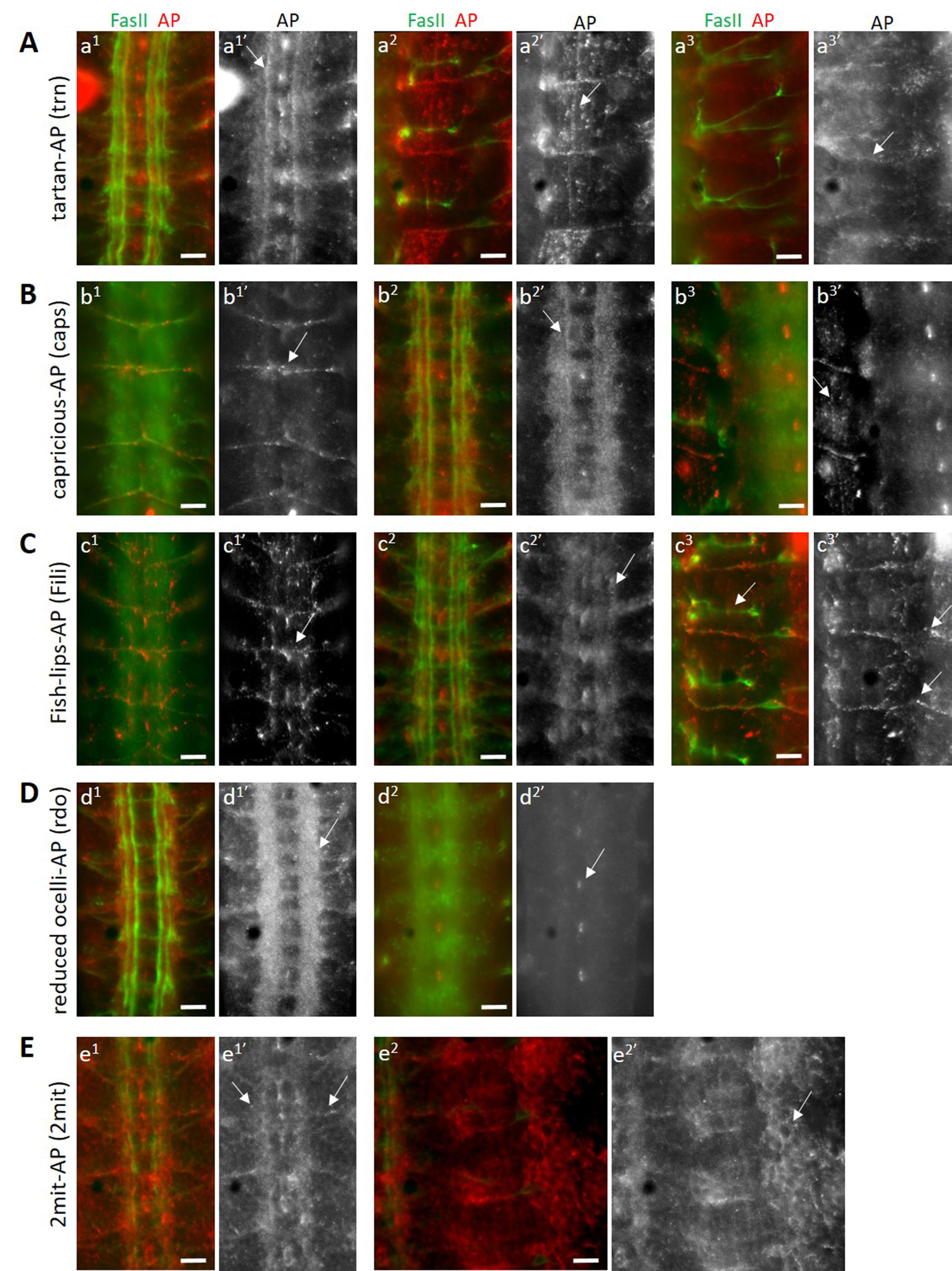
Tartan (trn) and Capricious (caps) are two closely-related eLRR proteins with known functions in embryonic motor axon guidance and the innervation of antennal lobe glomeruli by olfactory sensory axons (Kurusu et al. 2008; Hong et al. 2009). Studies of trn and caps single and double mutants suggest that the two genetically interact and may function through a common binding partner (Milan et al. 2005; Kurusu et al. 2008). Tartan may be a substrate for the receptor tyrosine phosphatase Ptp52F (Bugga et al. 2009). We stained wild-type live-dissected stage 16 Drosophila embryos with trn-AP and caps-AP fusion proteins separately, and found distinct as well as overlapping staining patterns for both fusion proteins. Both trn-AP and caps-AP bind to longitudinal axons in the ventral nerve cord (VNC), with stronger binding seen in one particular axon bundle close to the midline (arrows, a1’ and b2’). Both also show binding to muscles (arrows, a2’ and b3’), indicating that they interact with a binding partner expressed on the surface of muscles. trn-AP shows binding to a subset of sensory neurons (arrow, a3’), which caps-AP does not. In addition, caps-AP binds to the transverse nerve, which emanates from the midline and is located on the dorsal side of the VNC (arrow, b1’).

Fish-lips (Fili) is an eLRR with roles in the regulation of apoptosis (Adachi-Yamada et al. 2005) and olfactory receptor neuron (ORN) targeting in the antennal lobe (Xie et al. 2019). It is expressed at moderately-high levels during embryonic stages 12 – 17 and during 24 – 48 hours after puparium formation (modENCODE Temporal Expression Data, FlyBase). These developmental stages correspond to peak synaptogenesis times, implying a developmental role of Fili in regulating synaptogenesis. Thus, identification of binding partners of Fili is crucial to understand its roles in CNS development. Staining of wild-type stage 16 embryos with Fili-AP fusion protein shows a restricted binding pattern in the CNS, indicating a similar restricted expression pattern of its binding partners. It binds to a set of dorsal midline neurons (arrow, c1’) and a subset of longitudinal axons in the VNC (arrow, c2’). A subset of midline cells, putatively glial cells are also labeled with Fili-AP. Strong binding is seen to the transverse nerve in the VNC (c1) and in the periphery (arrow, c3’), while no labeling is seen to the SNa in the same focal plane (arrow, c3).

Reduced ocelli (rdo) is a gene that regulates ocelli development (Caldwell et al. 2007) and encodes an eLRR protein of unknown function. Caldwell et al. 2007 showed a broad expression pattern of the encoded protein in the adult nervous system. We performed staining of wild-type stage 16 embryos with rdo-AP fusion protein and found a very strong binding signal in the longitudinal and commissural axons of the VNC (arrow, d1’). This binding was limited to the VNC, and no binding was observed to the muscles (data not shown), indicating that the eLRR encoded by rdo interacts with neuronal-specific ligands. We also observed binding in a subset of midline glial cells in the VNC (arrow, d2’).

2mit is another gene encoding a putative eLRR and is expressed in the developing nervous system. It has a putative role in regulating short-term memory (Baggio et al. 2013). No other information is known about this eLRR. We stained wild-type stage 16 embryos with 2mit-AP fusion protein and saw a wide pattern of binding by this fusion protein, unlike the other restricted patterns observed above. Both longitudinal, commissural as well as exiting motor axons in the VNC are labeled by 2mit-AP (arrows, e1’). Moreover, we observed a pan-cellular pattern of labeling in the periphery as well as in the VNC, where 2-mit-AP binding signal is seen on the surface of cells, resulting in a cell-membrane staining pattern (arrow, e2’). This implies that the eLRR encoded by 2mit is capable of interacting with ligands expressed on neuronal as well as non-neuronal cell types.

These binding patterns provide clues to the expression patterns of proteins that these eLRRs might interact with to regulate various developmental processes.

Reagents

y1 w1 (FlyBase ID FBst0001495)

References

Adachi-Yamada, T., T. Harumoto, K. Sakurai, R. Ueda, K. Saigo et al., 2005 Wing-to-Leg homeosis by spineless causes apoptosis regulated by Fish-lips, a novel leucine-rich repeat transmembrane protein. Mol Cell Biol 25: 3140-3150.
PubMed
Baggio, F., A. Bozzato, C. Benna, E. Leonardi, O. Romoli et al., 2013 2mit, an intronic gene of Drosophila melanogaster timeless2, is involved in behavioral plasticity. PLoS One 8: e76351.
PubMed
Bali, N., H. K. Lee and K. Zinn, 2016 Live Staining of Drosophila Embryos with RPTP Fusion Proteins to Detect and Characterize Expression of Cell-Surface RPTP Ligands. Methods Mol Biol 1447: 373-384.
PubMed
Bali, N., H.K. Lee and K. Zinn, 2019 Identification of four Drosophila Toll-related proteins as ligands for the PTP69D receptor tyrosine phosphatase. microPublication Biology 10.17912/micropub.biology.000159.
10.17912/micropub.biology.000159
Brose, K., K. S. Bland, K. H. Wang, D. Arnott, W. Henzel et al., 1999 Slit proteins bind Robo receptors and have an evolutionarily conserved role in repulsive axon guidance. Cell 96: 795-806.
PubMed
Bugga, L., A. Ratnaparkhi and K. Zinn, 2009 The cell surface receptor Tartan is a potential in vivo substrate for the receptor tyrosine phosphatase Ptp52F. Mol Cell Biol 29: 3390-3400.
PubMed
Caldwell, J. C., S. K. Fineberg and D. F. Eberl, 2007 reduced ocelli encodes the leucine rich repeat protein Pray For Elves in Drosophila melanogaster. Fly (Austin) 1: 146-152.
PubMed
de Wit, J., W. Hong, L. Luo and A. Ghosh, 2011 Role of leucine-rich repeat proteins in the development and function of neural circuits. Annu Rev Cell Dev Biol 27: 697-729.
PubMed
Dickson, B. J., and G. F. Gilestro, 2006 Regulation of commissural axon pathfinding by slit and its Robo receptors. Annu Rev Cell Dev Biol 22: 651-675.
PubMed
Dolan, J., K. Walshe, S. Alsbury, K. Hokamp, S. O'Keeffe et al., 2007 The extracellular leucine-rich repeat superfamily; a comparative survey and analysis of evolutionary relationships and expression patterns. BMC Genomics 8: 320.
PubMed
Fox, A. N., and K. Zinn, 2005 The heparan sulfate proteoglycan syndecan is an in vivo ligand for the Drosophila LAR receptor tyrosine phosphatase. Curr Biol 15: 1701-1711.
PubMed
Hong, W., H. Zhu, C. J. Potter, G. Barsh, M. Kurusu et al., 2009 Leucine-rich repeat transmembrane proteins instruct discrete dendrite targeting in an olfactory map. Nat Neurosci 12: 1542-1550.
PubMed
Kurusu, M., A. Cording, M. Taniguchi, K. Menon, E. Suzuki et al., 2008 A screen of cell-surface molecules identifies leucine-rich repeat proteins as key mediators of synaptic target selection. Neuron 59: 972-985.
PubMed
Lauren, J., M. S. Airaksinen, M. Saarma and T. Timmusk, 2003 A novel gene family encoding leucine-rich repeat transmembrane proteins differentially expressed in the nervous system. Genomics 81: 411-421.
PubMed
Lee, H. K., A. Cording, J. Vielmetter and K. Zinn, 2013 Interactions between a receptor tyrosine phosphatase and a cell surface ligand regulate axon guidance and glial-neuronal communication. Neuron 78: 813-826.
PubMed
Lee, H. K., A. P. Wright and K. Zinn, 2009 Live dissection of Drosophila embryos: streamlined methods for screening mutant collections by antibody staining. J Vis Exp.
PubMed
Milan, M., L. Perez and S. M. Cohen, 2005 Boundary formation in the Drosophila wing: functional dissection of Capricious and Tartan. Dev Dyn 233: 804-810.
PubMed
Ozkan, E., R. A. Carrillo, C. L. Eastman, R. Weiszmann, D. Waghray et al., 2013 An extracellular interactome of immunoglobulin and LRR proteins reveals receptor-ligand networks. Cell 154: 228-239.
PubMed
Xie, Q., B. Wu, J. Li, C. Xu, H. Li et al., 2019 Transsynaptic Fish-lips signaling prevents misconnections between nonsynaptic partner olfactory neurons. Proc Natl Acad Sci U S A 116:16068-16073.
PubMed

Funding

This work was supported by National Institutes of Health grant R37 NS28182 to K.Z. and by Gordon Ross Postdoctoral Fellowship to N.B.

Author Contributions

Namrata Bali: Investigation, Writing - original draft, Writing - review and editing
Kai Zinn: Conceptualization, Funding acquisition, Supervision, Writing - review and editing.

Reviewed By

Liqun Luo and Steven Marygold

History

Received: December 6, 2019
Accepted: December 16, 2019
Published: December 18, 2019

Copyright

© 2019 by the authors. This is an open-access article distributed under the terms of the Creative Commons Attribution 4.0 International (CC BY 4.0) License, which permits unrestricted use, distribution, and reproduction in any medium, provided the original author and source are credited.

Citation

Bali, N; Zinn, K (2019). Visualization of binding patterns for five Leucine-rich repeat proteins in the Drosophila embryo. microPublication Biology. 10.17912/micropub.biology.000199.
Download: RIS BibTeX
microPublication Biology is published by
1200 E. California Blvd. MC 1-43 Pasadena, CA 91125
The microPublication project is supported by
The National Institute of Health -- Grant #: 1U01LM012672-01
microPublication Biology:ISSN: 2578-9430