microPublication

Get Your Data Out, Be Cited

  • About
    • Editorial Policies
      • Editorial Staff
      • Editorial Board
      • Criteria For Publication
      • Publishing Information
      • Data Sharing Policy
    • For Authors
      • Preparation And Submission Of A Manuscript
      • Peer Review Process
      • Following Acceptance
      • Appeals
    • For Reviewers
    • Why micropublish?
  • Submit a microPublication
  • Journals
    • microPublication Biology
      • Editorial Board
  • microPublications
    • Biology
      • Species
        • Arabidopsis
        • C. elegans
        • D. discoideum
        • Drosophila
        • Human
        • Mouse
        • S. cerevisiae
        • S. pombe
        • Xenopus
        • Zebrafish
      • Categories
        • Phenotype Data
        • Methods
        • Expression Data
        • Genotype Data
        • Integrations
        • Genetic Screens
        • Models of Human Disease
        • Software
        • Interaction data
        • Database Updates
        • Electrophysiology Data
        • Phylogenetic Data
        • Science and Society
        • Biochemistry
  • Contact
  • More
    • Archives
    • FAQs
    • Newsletter
microPublication / Biology / Regulation of sleep by KIN-29...
Regulation of sleep by KIN-29 is not developmental
Jeremy J. Grubbs1, Alexander M. van der Linden2 and David M. Raizen1
1Department of Neurology, Perelman School of Medicine, University of Pennsylvania, Philadelphia, Pennsylvania 19104
2Department of Biology, University of Nevada, Reno, Reno, Nevada 89557
Correspondence to: Jeremy J. Grubbs (jeremy.fodor@pennmedicine.upenn.edu)
Figure 1: Regulation of sleep by KIN-29 is not developmental. A) Schematic of the CRISPR/Cas9 strain construction with the DEGRON and GFP-tags inserted at the end of the kin-29 open reading frame. For panels B, D, E, G and H, the average movement quiescence in minutes ± SEM are displayed following exposure to 1500 J/m2 UVC irradiation. (+) indicates that 2 mM auxin was present during development and analysis. B) UVC–SIS is similar to wild type (WT) in auxin–exposed tir1::mRuby and kin-29::degron::GFP animals, and in tir1::mRuby; kin-29::degron::GFP (KIN-29 AID) transgenic animals in the absence of auxin. C) An image of a KIN-29 AID L4 stage animal. GFP is expressed in head neurons (white arrow) and in the intestine (white arrow head) consistent with previous reports (Lanjuin and Sengupta 2002). tir1::mRuby is expressed ubiquitously, as reported (Zhang et al. 2015). D) UVC–SIS is impaired in KIN-29 AID animals exposed to auxin from the time of hatching. E) The UVC–SIS impairment produced by continuous auxin–induced degradation of KIN-29 is similar to that of kin-29(oy38) null mutants. F) Images of adult KIN-29 AID animals never exposed to auxin (top left), exposed to auxin since hatching (top right), exposed to auxin for 4 hours as adults (bottom left), and removed from auxin for 12 hours starting at the L4 stage after exposure from hatching (bottom right). White arrows point to head neurons (green), which are visible only in the absence of auxin. Images were captured with identical camera and microscope settings. G) KIN-29 AID animals show a similar defect in UVC–SIS whether exposed to auxin at hatch or from the adult stage for four hours. H) UVC–SIS in KIN-29 AID animals on auxin from hatching through adulthood (+) or until the L4 stage, and examined 12 hours later (-12 hours). Reduction of auxin-induced degradation of KIN-29 during adulthood partially restores SIS behavior. ns = not significant, *P<0.05, **P<0.01, ***P<0.001, ****P< 0.0001, two-way ANOVA or mixed-model analysis with Tukey correction for multiple comparisons. Comparisons are made against wild-type (WT) unless otherwise indicated.

Description

Sleep is intertwined with metabolic function in vertebrates (Tsuneki et al. 2016; Herrera et al. 2017; Aalling et al. 2018; Wilms et al. 2018) and invertebrates (Kempf et al. 2019; Ki and Lim 2019; Yurgel et al. 2019; Grubbs et al. 2020), but the molecular underpinnings of this connection are not well understood. We recently reported that the salt inducible kinase (SIK) homolog KIN-29 is required in a subset of sensory neurons for the metabolic regulation of sleep in Caenorhabditis elegans (Grubbs et al. 2020). However, since our genetic manipulations made use of mutations that were present throughout the life of the animal, it is possible that the sleep defect of kin-29 mutants reflects a requirement for KIN-29 activity during development rather than during sleep. Indeed, because kin-29 is expressed throughout development as well as adulthood (Maduzia et al. 2005), and kin-29 mutants show altered expression of targets influential in larval development (Van Der Linden et al. 2008), a role for kin-29 during development remained plausible. Distinguishing a developmental early role from a role during the time of sleep is important for constraining models for how KIN-29 regulates sleep.

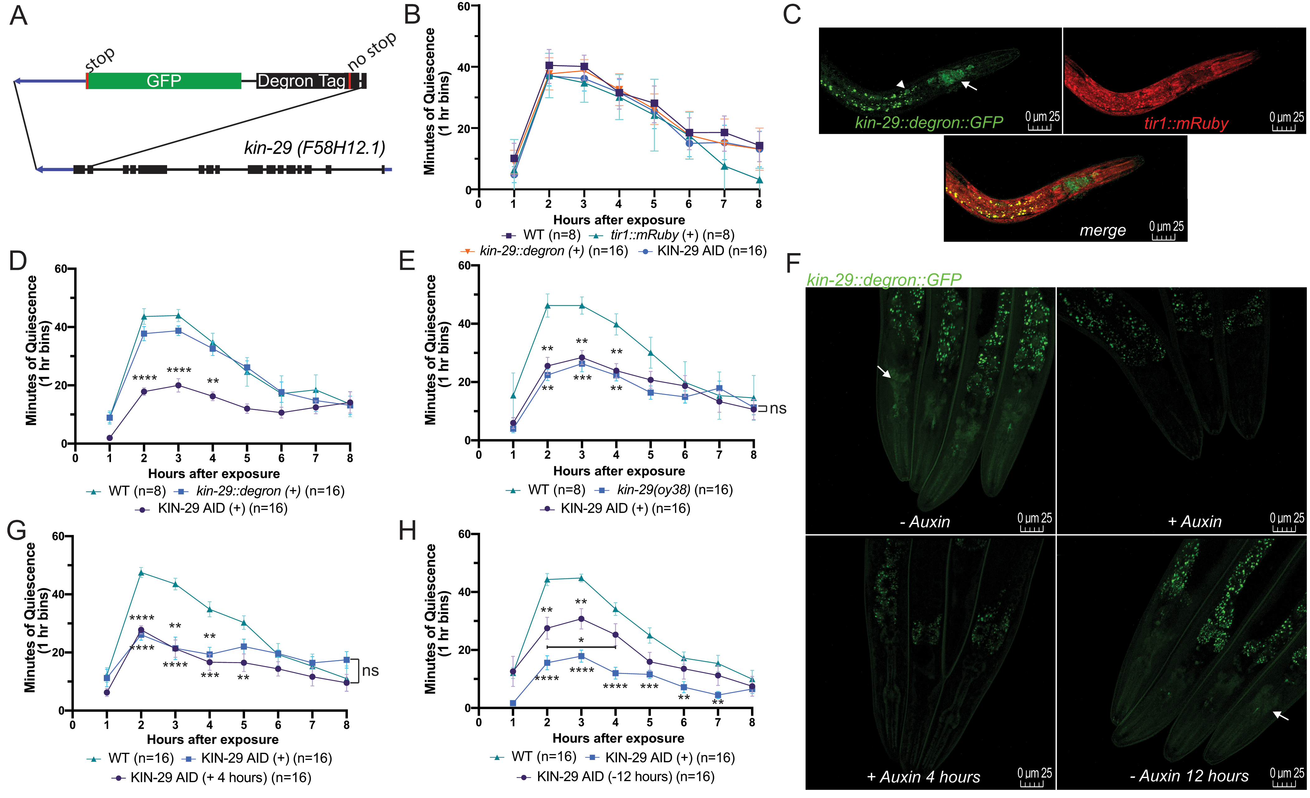
To determine the KIN-29 time window of action, we depleted the KIN-29 protein in a temporally-controlled fashion. We used CRISPR/Cas9 genome editing to introduce a DEGRON fused to GFP just before the kin-29 stop codon (Fig. 1A). After confirming successful editing by Sanger sequencing, we crossed the kin-29::degron::GFP into the previously generated Peft-3::TIR1::mRuby strain (Zhang et al. 2015) to allow for rapid and reversible degradation of KIN-29 when exposed to the phytohormone auxin (Ruegger et al. 1998; Gray et al. 1999). We examined stress-induced sleep (SIS) following exposure to UVC-irradiation (Debardeleben et al. 2017) in the first day of adulthood. We first verified that auxin exposure did not disrupt SIS in the parental strains, and that the progeny containing both transgenes, from here on referred to as KIN-29 Auxin-Inducible Degron (KIN-29 AID) animals, show normal SIS in the absence of auxin (Fig. 1B). We also confirmed the expression of both transgenes in KIN-29 AID animals (Fig. 1C). We then measured SIS in KIN-29 AID transgenic worms cultivated on agar containing auxin (2 mM) from the time of hatching. KIN-29 AID transgenic animals cultivated on auxin throughout development mimicked the kin-29(oy38) null mutant SIS phenotype (Fig. 1D-E). Such animals also showed undetectable expression of endogenous GFP-tagged KIN-29 (Fig. 1F).

To determine if KIN-29’s regulation of sleep has a developmental component, we used two paradigms. First, we cultivated young adult animals for four hours on agar containing auxin (2 mM). We then administered a UVC treatment (1500 J/m2) and recorded movement quiescence of animals in the continued presence of auxin. Four hours in the presence of auxin at the young adult stage was sufficient to reduce GFP-tagged KIN-29 expression (Fig. 1F), and was able to reduce SIS to a similar extent as that seen in animals cultivated on auxin their whole life (Fig. 1F-G). Second, to restore KIN-29 function to adult animals, we cultivated KIN-29 AID transgenic animals in the presence of auxin since hatching, but then removed them from auxin-containing plates at the L4 stage. We waited twelve hours before assessing SIS using UVC-irradiation treatment. These animals showed significantly increased movement quiescence in comparison to animals that were cultured their whole life on auxin, although these quiescence levels were lower than the quiescence phenotype of wt animals (Fig. 1H). Consistent with these intermediate behavioral results, expression of GFP-tagged KIN-29 in these animals was intermediate between that of animals never exposed to auxin, and that of animals exposed to auxin throughout life (Fig. 1F). Our results are consistent with previous observations that even after 24 hours off of auxin, expression of a DEGRON-tagged protein may not fully normalize (Zhang et al. 2015).

In summary, our results demonstrate that KIN-29’s function in SIS is not developmental. Rather, these findings suggest that kin-29 functions at the time of sleep. These results fit with our proposed model of kin-29 acting as a real-time energy sensor in sensory neurons to promote sleep (Grubbs et al. 2020). Based on this analysis, we propose that the sleep-regulatory effects of SIK3 in mammals (Funato et al. 2016) are also unlikely to be developmental.

Methods

Request a detailed protocol

Worm maintenance and strains

Worms were cultivated on the agar surface of 5.5 cm diameter Petri dishes filled with 11.5 mL of NGM agar. They were fed the Escherichia coli strain DA837 (Davis et al. 1995) and grown at 20°C. All experiments were performed on hermaphrodites. The wild-type strain used was N2, variety Bristol (Brenner 1974). Auxin experiments were carried out as described in (Zhang et al. 2015) using 2 mM auxin (Alfa Aesar #A10556). Strains available from the CGC are N2 and CA1200. Strains available from our lab are PHX1777 kin-29(syb1777[kin-29::degron::gfp]) and NQ1295 (ieSi57[Peft-3::tir1::mRuby]; kin-29(syb1777[kin-29::degron::gfp]).

CRISPR/Cas9 genome editing and crosses

CRISPR/Cas9-mediated genome editing was performed by SunyBiotech (China). The oligonucleotide sequence used for PCR and Sequencing were (Sense) 5’CATTGTGTGCACATTACCGT and (Antisense) 5’AATCAGCTAGCACAGGCTCT. To generate the strain NQ1295, PHX1777 males were crossed with CA1200 hermaphrodites, with the red and green fluorescent phenotypes followed using a Leica SP8 confocal microscope. We found a regular epifluorescence compound microscope not sufficiently sensitive to detect the fluorescence in these strains, which was very dim.

Ultraviolet light exposure and movement quiescence recordings

Movement quiescence was quantified using machine vision with images recorded using the 48-well WorMotel (Churgin et al. 2019). Briefly, animals were selected as L4 larvae 12-16 hours prior to behavioral recording. All 48 wells were filled with NGM agar with or without 2 mM auxin, which was diluted from a 400 mM stock as described in (Zhang et al. 2015), and a thin layer of bacteria was spread over the agar surface using a platinum wire. Animals were loaded individually as adults into WorMotel wells and then treated using a Spectrolinker XL-1000 crosslinker with 1500 J/m2 of UVC (254 nm) radiation. The WorMotel was placed under a DMK 23GP031 camera (The Imaging Source) for recording. Images were captured every 10 seconds for 8 hours. Images collected of the animals on the WorMotel were analyzed with custom MatLab scripts (Churgin et al. 2019). The quantification of movement was determined via pixel subtraction and an animal was considered quiescent if there was no pixel change between images.

Confocal imaging

Animals were placed in a 1ml droplet of M9 buffer and immobilized on a 2% agarose pad containing 25mM levamisole. Images were taken using a Leica SP8 confocal microscope as Z-stacks with 0.76 microns between images in a stack and a total of 36 images per condition taken. The powers of the 488 nm and 552 nm lasers were set to 2.5% and 2% respectively. Z-projection was performed in FIJI (Version 2.0.0).

Acknowledgments

We would like to thank SunyBiotech for generation of the kin-29::degron::GFP tagged (PHX1777) strain.

References

Aalling, N. N., Nedergaard, M., and DiNuzzo, M. (2018). Cerebral Metabolic Changes During Sleep. Curr Neurol Neurosci Rep, 18(9), 57.
PubMed
Brenner, S. (1974). The genetics of Caenorhabditis elegans. Genetics, 77(1), 71-94.
PubMed
Churgin, M. A., Szuperak, M., Davis, K. C., Raizen, D. M., Fang-Yen, C., and Kayser, M. S. (2019). Quantitative imaging of sleep behavior in Caenorhabditis elegans and larval Drosophila melanogaster. Nature Protocols, 14(5), 1455-1488.
PubMed
Davis, M. W., Somerville, D., Lee, R. Y., Lockery, S., Avery, L., and Fambrough, D. M. (1995). Mutations in the Caenorhabditis elegans Na,K-ATPase alpha-subunit gene, eat-6, disrupt excitable cell function. J Neurosci, 15(12), 8408-8418.
PubMed
DeBardeleben, H. K., Lopes, L. E., Nessel, M. P., and Raizen, D. M. (2017). Stress-Induced Sleep After Exposure to Ultraviolet Light Is Promoted by p53 in Caenorhabditis elegans. Genetics, 207(2), 571-582.
PubMed
Funato, H., Miyoshi, C., Fujiyama, T., Kanda, T., Sato, M., Wang, Z., Ma, J., Nakane, S., Tomita, J., Ikkyu, A., Kakizaki, M., Hotta-Hirashima, N., Kanno, S., Komiya, H., Asano, F., Honda, T., Kim, S. J., Harano, K., Muramoto, H., Yonezawa, T., Mizuno, S., Miyazaki, S., Connor, L., Kumar, V., Miura, I., Suzuki, T., Watanabe, A., Abe, M., Sugiyama, F., Takahashi, S., Sakimura, K., Hayashi, Y., Liu, Q., Kume, K., Wakana, S., Takahashi, J. S. and Yanagisawa, M. (2016). Forward-genetics analysis of sleep in randomly mutagenized mice. Nature, 539(7629), 378-383.
PubMed
Gray, W. M., del Pozo, J. C., Walker, L., Hobbie, L., Risseeuw, E., Banks, T., Crosby, W.L., Yang, M., Ma H., and Estelle, M. (1999). Identification of an SCF ubiquitin–ligase complex required for auxin response in Arabidopsis thaliana. Genes & Development, 13(13), 1678-1691.
PubMed
Grubbs, J. J., Lopes, L. E., van der Linden, A. M., and Raizen, D. M. (2020). A salt-induced kinase is required for the metabolic regulation of sleep. PLOS Biology, 18(4), e3000220.
PubMed
Herrera, C. G., Ponomarenko, A., Korotkova, T., Burdakov, D., and Adamantidis, A. (2017). Sleep & metabolism: The multitasking ability of lateral hypothalamic inhibitory circuitries. Frontiers in Neuroendocrinology, 44, 27-34.
PubMed
Kempf, A., Song, S. M., Talbot, C. B., and Miesenböck, G. (2019). A potassium channel β-subunit couples mitochondrial electron transport to sleep. Nature, 568(7751), 230-234.
PubMed
Ki, Y., and Lim, C. (2019). Sleep-promoting effects of threonine link amino acid metabolism in Drosophila neuron to GABAergic control of sleep drive. Elife, 8.
PubMed
Lanjuin, A., and Sengupta, P. (2002). Regulation of chemosensory receptor expression and sensory signaling by the KIN-29 Ser/Thr kinase. Neuron, 33(3), 369-381.
PubMed
Maduzia, L. L., Roberts, A. F., Wang, H., Lin, X., Chin, L. J., Zimmerman, C. M., Cohen, S., Feng, X. H. and Padgett, R. W. (2005). C. elegans serine-threonine kinase KIN-29 modulates TGFbeta signaling and regulates body size formation. BMC Dev Biol. 5, 8.
PubMed
Ruegger, M., Dewey, E., Gray, W. M., Hobbie, L., Turner, J., and Estelle, M. (1998). The TIR1 protein of Arabidopsis functions in auxin response and is related to human SKP2 and yeast Grr1p. Genes & Development, 12(2), 198-207.
PubMed
Tsuneki, H., Sasaoka, T., and Sakurai, T. (2016). Sleep Control, GPCRs, and Glucose Metabolism. Trends in Endocrinology & Metabolism, 27(9), 633-642.
PubMed
van der Linden, A. M., Wiener, S., You, Y. J., Kim, K., Avery, L., and Sengupta, P. (2008). The EGL-4 PKG acts with KIN-29 salt-inducible kinase and protein kinase A to regulate chemoreceptor gene expression and sensory behaviors in Caenorhabditis elegans. Genetics, 180(3), 1475-1491.
PubMed
Wilms, B., Leineweber, E. M., Mölle, M., Chamorro, R., Pommerenke, C., Salinas-Riester, G., Sina, C., Lehnert, H., Oster, H., and Schmid, S. M. (2018). Sleep Loss Disrupts Morning-to-Evening Differences in Human White Adipose Tissue Transcriptome. The Journal of Clinical Endocrinology & Metabolism, 104(5), 1687-1696.
PubMed
Yurgel, M. E., Kakad, P., Zandawala, M., Nassel, D. R., Godenschwege, T. A., and Keene, A. C. (2019). A single pair of leucokinin neurons are modulated by feeding state and regulate sleep-metabolism interactions. PLoS Biol, 17(2), e2006409.
PubMed
Zhang, L., Ward, J. D., Cheng, Z., and Dernburg, A. F. (2015). The auxin-inducible degradation (AID) system enables versatile conditional protein depletion in C. elegans. Development, 142(24), 4374-4384.
PubMed

Funding

This work was supported by the National Institutes of Health grant R01NS107969. The CA1200 strain was provided by the Caenorhabditis Genetics Center, which is funded by the NIH office of Research Infrastructure Programs (P40 OD010440).

Author Contributions

Jeremy J. Grubbs: Formal analysis, Methodology, Investigation, Writing - original draft, Writing - review and editing
Alexander M. van der Linden: Funding acquisition, Resources, Writing - review and editing
David M. Raizen: Funding acquisition, Writing - review and editing, Supervision.

Reviewed By

Cheryl Van Buskirk

History

Received: April 28, 2020
Revision received: May 6, 2020
Accepted: May 7, 2020
Published: May 7, 2020

Copyright

© 2020 by the authors. This is an open-access article distributed under the terms of the Creative Commons Attribution 4.0 International (CC BY 4.0) License, which permits unrestricted use, distribution, and reproduction in any medium, provided the original author and source are credited.

Citation

Grubbs, JJ; van der Linden, AM; Raizen, DM (2020). Regulation of sleep by KIN-29 is not developmental. microPublication Biology. 10.17912/micropub.biology.000247.
Download: RIS BibTeX
microPublication Biology is published by
1200 E. California Blvd. MC 1-43 Pasadena, CA 91125
The microPublication project is supported by
The National Institute of Health -- Grant #: 1U01LM012672-01
microPublication Biology:ISSN: 2578-9430