Description
VWA8 proteins, named for von Willebrand factor A (VWA) domain containing 8, are conserved from worm to mammals (Whittaker & Hynes, 2002). In mouse, vwa8 gene produces long and short protein isoforms (VWA8a and VWA8b), and in rat livers VWA8a has been shown to localize to the matrix side of inner mitochondrial membrane (Luo et al., 2020).
C. elegans vwa-8 is also predicted to produce two major protein isoforms. The short isoform (974 AA) shares identical AA sequences as the long isoform (1804 AA). The two isoforms contain an MTS (mitochondrial targeting sequence) at the N terminus, followed by three AAA ATPase domains, which are associated with diverse cellular activities; and the long isoform contains a VWA domain at the C terminus.
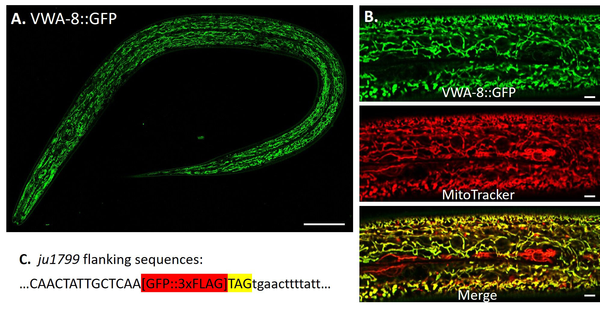
To determine the endogenous expression pattern of C. elegans VWA-8, we generated a GFP knock-in allele, ju1799, in which GFP was in-frame fused at the C terminus to label the full-length protein specifically. The endogenous VWA-8::GFP was expressed in mitochondria of hypodermis, intestine and muscle, but was not detectable in neurons (Fig 1A). In hypodermis, VWA-8::GFP was colocalized with MitoTracker Red, a mitochondria marker (Fig 1B).
Methods
Request a detailed protocolCRISPR-mediated GFP knock-in: A GFP::3xFLAG tag was inserted right before the STOP codon of vwa-8 long isoform, following a CRISPR-Cas9 protocol (Dickinson, Pani, Heppert, Higgins, & Goldstein, 2015). We designed a subgenomic RNA (sgRNA): AGCAATAGTTGATGAGAAAA targeting the stop codon of vwa-8. We injected 50 ng/µl of vwa-8 sgRNA, 10 ng/µl of homology arm repair template, 2.5 ng/µl Pmyo-2::mCherry and 5 ng/µl of Pmyo-3::mCherry into wild type worms. 3 days after injection hygromycin was added to the plates to kill the untransformed F1 animals. On day 6 post-injection, we looked for candidate GFP knock-in animals which were L4/adult roller, survived hygromycin selection and without the mCherry extrachromosomal array markers. We then heat shocked 20 L1/L2 candidate knock-in worms at 34°C for 4 hours to remove the self-excising cassette. After that, the WT-looking worms were the final GFP knock-in animals. The GFP fluorescence was examined using compound microscopy. GFP insertion was confirmed by PCR using primers: 5’-GGGGCGGATGATGAGAAGTT-3’ and 5’-TGCTCTCGAACACCTTGCTT-3’.
MitoTracker Red CMXRos staining: Worms were soaked in 50 µl MitoTracker Red CMXRos (2.5 µM in M9 buffer; Invitrogen M7512) for 10 min at 20°C in the dark. Then worms were transferred to an OP50-seeded NGM plate and allowed to recover for 2 h at 20°C in the dark.
Imaging: Fluorescence images were collected using Zeiss LSM800 confocal microscopy. Worms were anesthetized with 2µM of levamisole.
Reagents
CZ27748 vwa-8::GFP::3xFLAG (ju1799) will be available at the CGC.
Acknowledgments
We thank Dr. Junxiang Zhou for advice on MitoTracker staining, and members of the Jin and Chisholm laboratories for valuable discussions. We acknowledge WormBase as an information resource.
References
Funding
This work was supported by NIH R01 NS093588 to AC and YJ.
Reviewed By
Iqbal HamzaHistory
Received: May 20, 2020Revision received: May 23, 2020
Accepted: May 24, 2020
Published: June 3, 2020
Copyright
© 2020 by the authors. This is an open-access article distributed under the terms of the Creative Commons Attribution 4.0 International (CC BY 4.0) License, which permits unrestricted use, distribution, and reproduction in any medium, provided the original author and source are credited.Citation
Zhu, M; Chisholm, AD; Jin, Y (2020). C. elegans VWA-8 is a mitochondrial protein. microPublication Biology. 10.17912/micropub.biology.000264.Download: RIS BibTeX


