Departamento de Fisiología, Biología Molecular y Celular, Facultad de Ciencias Exactas y Naturales, Universidad de Buenos Aires, Buenos Aires, Argentina
Instituto de Investigaciones Bioquímicas de Bahía Blanca (CONICET)
Departamento de Biología, Bioquímica y Farmacia, Universidad Nacional del Sur, Bahía Blanca, Argentina.
Consejo Nacional de Investigaciones Científicas y Técnicas
Instituto de Biociencias, Biotecnología y Biología traslacional, Departamento de Fisiología, Biología Molecular y Celular, Facultad de Ciencias Exactas y Naturales, Universidad de Buenos Aires, Buenos Aires, Argentina

Description
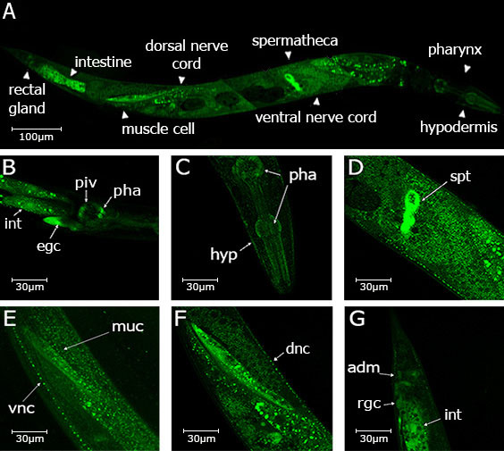
The endoplasmic reticulum (ER) uses an elaborate system called the ER quality control (QC) to monitor the proper folding of newly synthesized glycoproteins. The QC allows cells to differentiate between properly folded and misfolded proteins, allowing only those proteins which have acquired their native conformations to exit the ER and reach their final destinations. Alternatively, misfolded glycoproteins or incompletely formed glycoprotein complexes are translocated to the cytosol where they are finally degraded by proteasomes (Caramelo and Parodi 2007). The key element of this mechanism is the UDP-Glc: glycoprotein glucosyltransferase (UGGT) that functions as a folding sensor as it glucosylates exclusively those glycoproteins that have not acquired their native structures (Trombetta et al., 1989; Caramelo et al., 2003, 2004). Only vertebrates and Caenorhabditis genomes carry two uggt gene copies (uggt–1 and uggt–2) and phylogenetic inference showed that uggt genes went through independent duplications in Caenorhabditis and vertebrates. UGGT-1 retained canonical UGGT activity both in vertebrates and Caenorhabditis and vertebrate UGGT-2 underwent a specialization process. In Caenorhabditis, uggt-2 evolved by means of a putative neofunctionalization process in a non-redundant paralog and its biological function is still unknown (Caraballo et al., 2020; Buzzi et al.., 2011). Hence, UGGT-1 is the only protein engaged in monitoring the folding state of every glycoprotein in Caenorhabditis ER. To determine C. elegans UGGT-1’s body pattern expression we used fosmid recombineering technology (Tursun et al., 2009) to generate the Puggt-1::sl2::nls::gfp::unc-54 3’UTR transcriptional fusion reporter and established worm lines expressing this construct. UGGT-1 is expressed in the head, both in the pharynx, (corpus, isthmus and terminal bulb and buccal cavity) and in the pharyngeal intestinal valve. In the same image its expression is detected in the hypodermis and in the secretory gland (B and C). The somatic cells of the spermatheca express UGGT-1, but not the germline (D). Consistent with our previous findings (Buzzi et al.., 2011) UGGT-1 is widely expressed in the nervous system, both in ventral and dorsal nerve cords (E and F), as well as in the muscle cells as shown in (E-F) and in the anal depressor muscle (G). In the tail expression is also observed both in the rectal gland cell and the intestine.
Methods
Request a detailed protocolWorms of stable transgenic lines carrying the exaEx101 [Puggt-1::sl2::nls::gfp::unc-54 3’UTR] transcriptional fusion reporter were visualized by fluorescence confocal microscopy using an LSM510 Meta confocal microscope (Carl Zeiss, Oberkochen, Germany). Images were acquired with LSM software (Carl Zeiss) using a 20 x plan apochromat objective.
Acknowledgments
Armando Parodi and Mark Alkema
References
Funding
PIP 20080100567, National Research Council (Argentina)
Reviewed By
AnonymousHistory
Received: July 30, 2020Revision received: August 20, 2020
Accepted: August 24, 2020
Published: August 25, 2020
Copyright
© 2020 by the authors. This is an open-access article distributed under the terms of the Creative Commons Attribution 4.0 International (CC BY 4.0) License, which permits unrestricted use, distribution, and reproduction in any medium, provided the original author and source are credited.Citation
Buzzi, L; Segobia, VA; Rayes, D; Castro, OA (2020). The ER glycoprotein folding sensor UDP-Glc: glycoprotein glucosyltransferase is broadly expressed in C. elegans hermaphrodite. microPublication Biology. 10.17912/micropub.biology.000299.Download: RIS BibTeX
