microPublication

Get Your Data Out, Be Cited

  • About
    • Editorial Policies
      • Editorial Staff
      • Editorial Board
      • Criteria For Publication
      • Publishing Information
      • Data Sharing Policy
    • For Authors
      • Preparation And Submission Of A Manuscript
      • Peer Review Process
      • Following Acceptance
      • Appeals
    • For Reviewers
    • Why micropublish?
  • Submit a microPublication
  • Journals
    • microPublication Biology
      • Editorial Board
  • microPublications
    • Biology
      • Species
        • Arabidopsis
        • C. elegans
        • D. discoideum
        • Drosophila
        • Human
        • Mouse
        • S. cerevisiae
        • S. pombe
        • Xenopus
        • Zebrafish
      • Categories
        • Phenotype Data
        • Methods
        • Expression Data
        • Genotype Data
        • Integrations
        • Genetic Screens
        • Models of Human Disease
        • Software
        • Interaction data
        • Database Updates
        • Electrophysiology Data
        • Phylogenetic Data
        • Science and Society
        • Biochemistry
  • Contact
  • More
    • Archives
    • FAQs
    • Newsletter
microPublication / Biology / Endogenous expression and localization of...
Endogenous expression and localization of CAV-1::GFP in C. elegans
Dillon E. Sloan1 and Joshua N. Bembenek1
1University of Michigan
Correspondence to: Joshua N. Bembenek (bembenek@umich.edu)
Figure 1. Endogenous CAV-1::GFP: (A) Schematic of cav-1 edit (B-G) Single-plane images of CAV-1::GFP localization: at the bend of the gonad arm (B), in the -1 oocyte (C), in a prometaphase I embryo (D), in an anaphase I embryo (E), in a bean stage embryo (F), and in a fold-stage embryo (G). (H) CAV-1::GFP in adult worm. Numbers correspond with number of cells in embryo.

Description

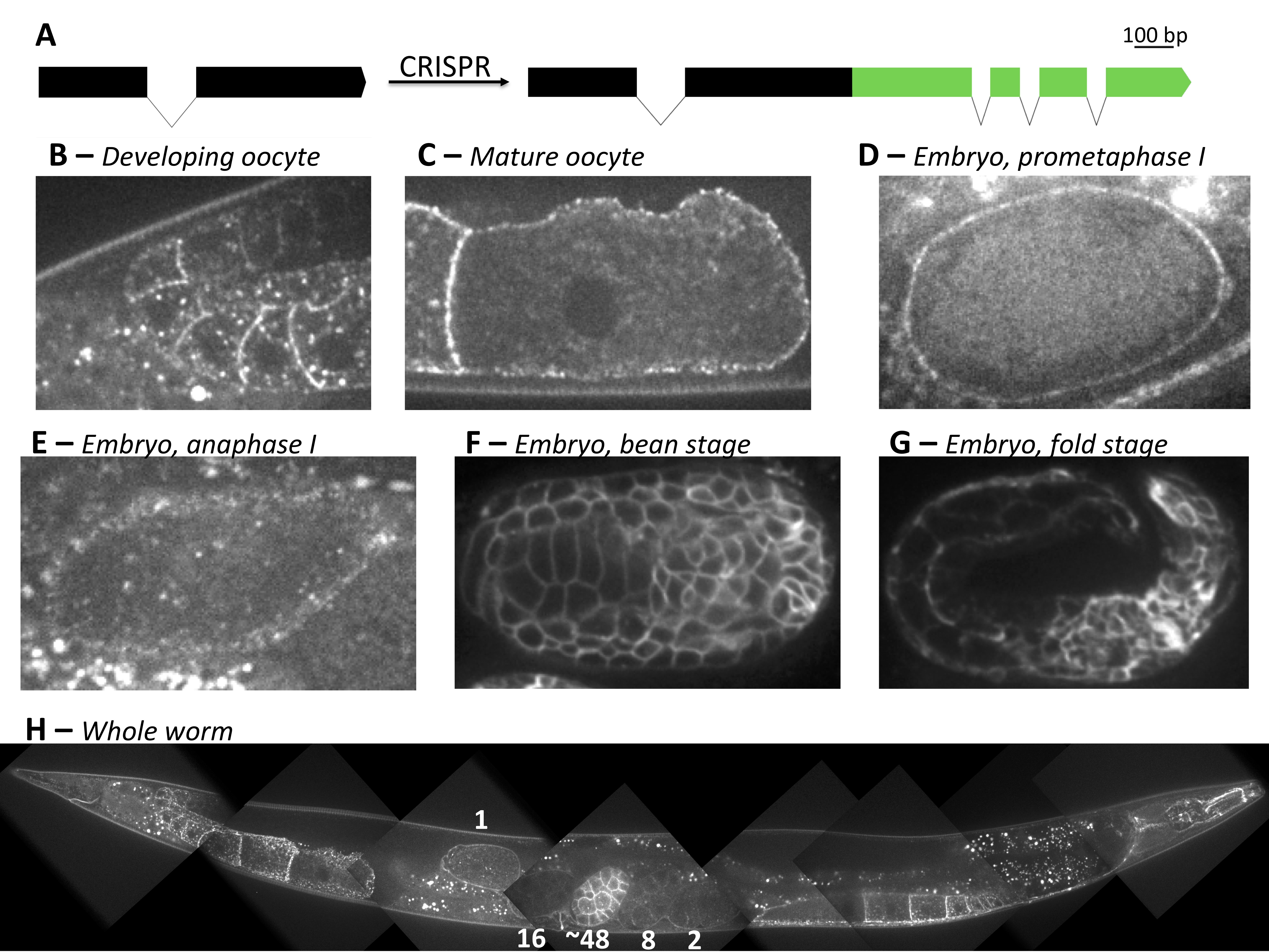
Caveolins are integral membrane proteins responsible for the formation of caveolae, invaginations of the plasma membrane linked to various disease states (Parton et al. 2020). In C. elegans, there are two caveolin proteins, CAV-1 and CAV-2. The cav-1 gene shares homology with all three mammalian caveolin genes (Tang et al. 1997). C. elegans CAV-1 protein does not appear to form caveolae, but a double-knockout mutant of cav-1 and cav-2 affects egg laying, and knockdown of cav-1 affects locomotion in a dynamin mutant background (Parker et al.. 2007, Kirkham et al. 2008, Sato et al. 2008). Based on exogenous expression, CAV-1::GFP is known to localize to cortical granules and the plasma membrane in oocytes and the early embryo, to the plasma membrane in later embryos, as well as to the neuromuscular system in larvae and adult worms (Sato et al. 2006, Bembenek et al. 2007, Parker et al. 2007).

Using CRISPR/Cas9, we generated an endogenous CAV-1::GFP fluorescent fusion strain, primarily to create a cortical granule marker (Figure 1A). Imaging endogenous CAV-1::GFP revealed similar patterns as previously reported in the germ line (Sato et al. 2006). However, unlike the pie-1 driven exogenous line, endogenous CAV-1::GFP was not clearly detected in the distal gonad until the bend region of the gonad arm, where it localized to the plasma membrane, and vesicle-shaped structures (Figure 1B). Oocytes had slightly higher expression showing enrichment at plasma membrane localization but faint cytoplasmic signal, and no obvious enrichment to vesicles (Figure 1C). After ovulation, CAV-1::GFP remained on the plasma membrane during meiosis I and was no longer detected by meiosis II (Figure 1D,E). In 5/5 anaphase I embryos, CAV-1::GFP was not significantly localized to cortical granules at a time when separase could be observed on vesicles. Overall, endogenous CAV-1::GFP signal is less intense in the germline than exogenous pie-1-driven lines. However, endogenous CAV-1::GFP signal became increasingly intense at the plasma membrane sometime after the 16-cell embryo stage (Figure 1H). The membrane signal remains intense through later stages of embryonic development (Figure 1F,G). This potentially indicates a dynamic role for cav-1 in embryonic development. Finally, adult somatic expression appeared to follow previously observed patterns (Figure 1H, Parker et al. 2007).

In summary, we have developed an endogenous CAV-1::GFP line by CRISPR, which can be of use to researchers interested in CAV-1.

Methods

Request a detailed protocol

CRISPR/Cas9 Gene editing

We followed the CRISPR/Cas9 protocol generated by Seydoux lab for C-terminal GFP tagging of the C. elegans cav-1 gene (Figure 1A, Paix et al. 2015). The repair template was amplified from the pDD282 plasmid. All guide RNAs and oligos were obtained commercially.

The primer sequences are listed below:

cav-1::GFP Forward (termed DS_027):

ACGGAATCAATCAAGAAACTACTGCTCCATGCGTCATGAGTAAAGGAGAAGAATTGTTCACTG

cav-1::GFP Reverse (termed DS_026):

AGTAAAATGAATTTGAGATAAATTAAATAAATTTACTTGTAGAGCTCGTCCATTCC

The crRNA sequence recognizing the spacer upstream of NGG is as follows:

(Termed DS_g003): AUUAAAUAAAUUUAGACGCA

Microscopy

Imaging was done as indicated in Bai et al. 2020. Embryos were staged using the localization of separase fused with a red fluorescent protein as well as embryo position and age in the animal.

Reagents

JAB192: cav-1(erb78[cav-1::GFP])

This strain is available upon request.

Acknowledgments

We thank members of the Bembenek and Csankovszki labs, especially Chris Turpin, for useful feedback.

References

Bai X., Melesse M., Sorensen Turpin C.G., Sloan D.E., Chen C.Y., Wang W.C., Lee P.Y., Simmons J.R., Nebenfuehr B., Mitchell D., Klebanow L.R., Mattson N., Betzig E., Chen B.C., Cheerambathur D., Bembenek J.N. (2020). Aurora B functions at the apical surface after specialized cytokinesis during morphogenesis in C. elegans. Development 147(1), dev181099.
PubMed
Bembenek, J.N., Richie, C.T., Squirrell, J.M., Campbell, J.M., Eliceiri, K.W., Poteryaev, D., Spang, A., Golden, A., and White, J.G. (2007). Cortical granule exocytosis in C. elegans is regulated by cell cycle components including separase. Development 134, 3837–3848.
PubMed
Kirkham, M., Nixon, S.J., Howes, M.T., Abi-Rached, L., Wakeham, D.E., Hanzal-Bayer, M., Ferguson, C., Hill, M.M., Fernandez-Rojo, M., Brown, D.A., Hancock, J.F., Brodsky, F.M., Parton, R.G. (2008). Evolutionary analysis and molecular dissection of caveola biogenesis. J. Cell Sci. 121, 2075–2086.
PubMed
Paix, A., Folkmann, A., Rasoloson, D., Seydoux, G. (2015). High efficiency, homology-directed genome editing in Caenorhabditis elegans using CRISPR–Cas9 ribonucleoprotein complexes. Genetics 201, 47–54.
PubMed
Parker, S., Peterkin, H. S., Baylis, H. A. (2007). Muscular dystrophy associated mutations in caveolin-1 induce neurotransmission and locomotion defects in Caenorhabditis elegans. Invert. Neurosci 7, 157-164.
PubMed
Parton R.G., McMahon K.A., Wu Y. (2020). Caveolae: Formation, dynamics, and function. Curr Opin Cell Biol 65, 8-16.
PubMed
Sato, K., Sato, M., Audhya, A., Oegema, K., Schweinsberg, P., and Grant, B. D. (2006). Dynamic regulation of caveolin-1 trafficking in the germ line and embryo of Caenorhabditis elegans. Mol. Biol. Cell 17, 3085-3094.
PubMed
Sato, M., Grant, B.D., Harada, A., Sato, K. (2008). Rab11 is required for synchronous secretion of chondroitin proteoglycans after fertilization in Caenorhabditis elegans. J Cell Sci 121, 3177–3186.
PubMed
Tang, Z., Okamoto, T., Boontrakulpoontawee, P., Katada, T., Otsuka, A. J., and Lisanti, M. P. (1997). Identification, sequence, and expression of an invertebrate caveolin gene family from the nematode Caenorhabditis elegans. Implications for the molecular evolution of mammalian caveolin genes. J. Biol. Chem. 272, 2437–2445.
PubMed

Funding

National Institutes of Health (R01 GM114471)

Author Contributions

Dillon E. Sloan: Data curation, Formal analysis, Investigation, Methodology, Writing - original draft, Writing - review and editing, Software
Joshua N. Bembenek: Conceptualization, Funding acquisition, Methodology, Project administration, Supervision, Validation, Writing - review and editing.

Reviewed By

Sara Olson

History

Received: August 6, 2020
Revision received: September 18, 2020
Accepted: September 18, 2020
Published: September 21, 2020

Copyright

© 2020 by the authors. This is an open-access article distributed under the terms of the Creative Commons Attribution 4.0 International (CC BY 4.0) License, which permits unrestricted use, distribution, and reproduction in any medium, provided the original author and source are credited.

Citation

Sloan, DE; Bembenek, JN (2020). Endogenous expression and localization of CAV-1::GFP in C. elegans. microPublication Biology. 10.17912/micropub.biology.000311.
Download: RIS BibTeX
microPublication Biology is published by
1200 E. California Blvd. MC 1-43 Pasadena, CA 91125
The microPublication project is supported by
The National Institute of Health -- Grant #: 1U01LM012672-01
microPublication Biology:ISSN: 2578-9430