microPublication

Get Your Data Out, Be Cited

  • About
    • Editorial Policies
      • Editorial Staff
      • Editorial Board
      • Criteria For Publication
      • Publishing Information
      • Data Sharing Policy
    • For Authors
      • Preparation And Submission Of A Manuscript
      • Peer Review Process
      • Following Acceptance
      • Appeals
    • For Reviewers
    • Why micropublish?
  • Submit a microPublication
  • Journals
    • microPublication Biology
      • Editorial Board
  • microPublications
    • Biology
      • Species
        • Arabidopsis
        • C. elegans
        • D. discoideum
        • Drosophila
        • Human
        • Mouse
        • S. cerevisiae
        • S. pombe
        • Xenopus
        • Zebrafish
      • Categories
        • Phenotype Data
        • Methods
        • Expression Data
        • Genotype Data
        • Integrations
        • Genetic Screens
        • Models of Human Disease
        • Software
        • Interaction data
        • Database Updates
        • Electrophysiology Data
        • Phylogenetic Data
        • Science and Society
        • Biochemistry
  • Contact
  • More
    • Archives
    • FAQs
    • Newsletter
microPublication / Biology / mNG-tagged mls-2 knock-in alleles in...
mNG-tagged mls-2 knock-in alleles in C. elegans
Rui Xiong1, Yi-Wen Hsieh1 and Chiou-Fen Chuang1,2
1Department of Biological Sciences, University of Illinois at Chicago
2Graduate Program in Neuroscience, University of Illinois at Chicago
Correspondence to: Chiou-Fen Chuang (chioufen.chuang@gmail.com)

Abstract

The Caenorhabditis elegans HMX/NKX MLS-2 transcription factor was previously shown to play sequential roles in AWC general identity and the stochastic AWCON/AWCOFF subtype choice during embryogenesis. Here we analyze the expression pattern of endogenous mls-2 during AWC development using mNeonGreen (mNG) knock-in strains. Similar to transgenic GFP::MLS-2, functional mNG::MLS-2 knock-in displayed nuclear localization in AWC precursor cells but was not observed in AWC during the later embryonic stage. These results suggest that the expression of mls-2 is below the detectable level and/or the stability of MLS-2 protein is very low in AWC cells.

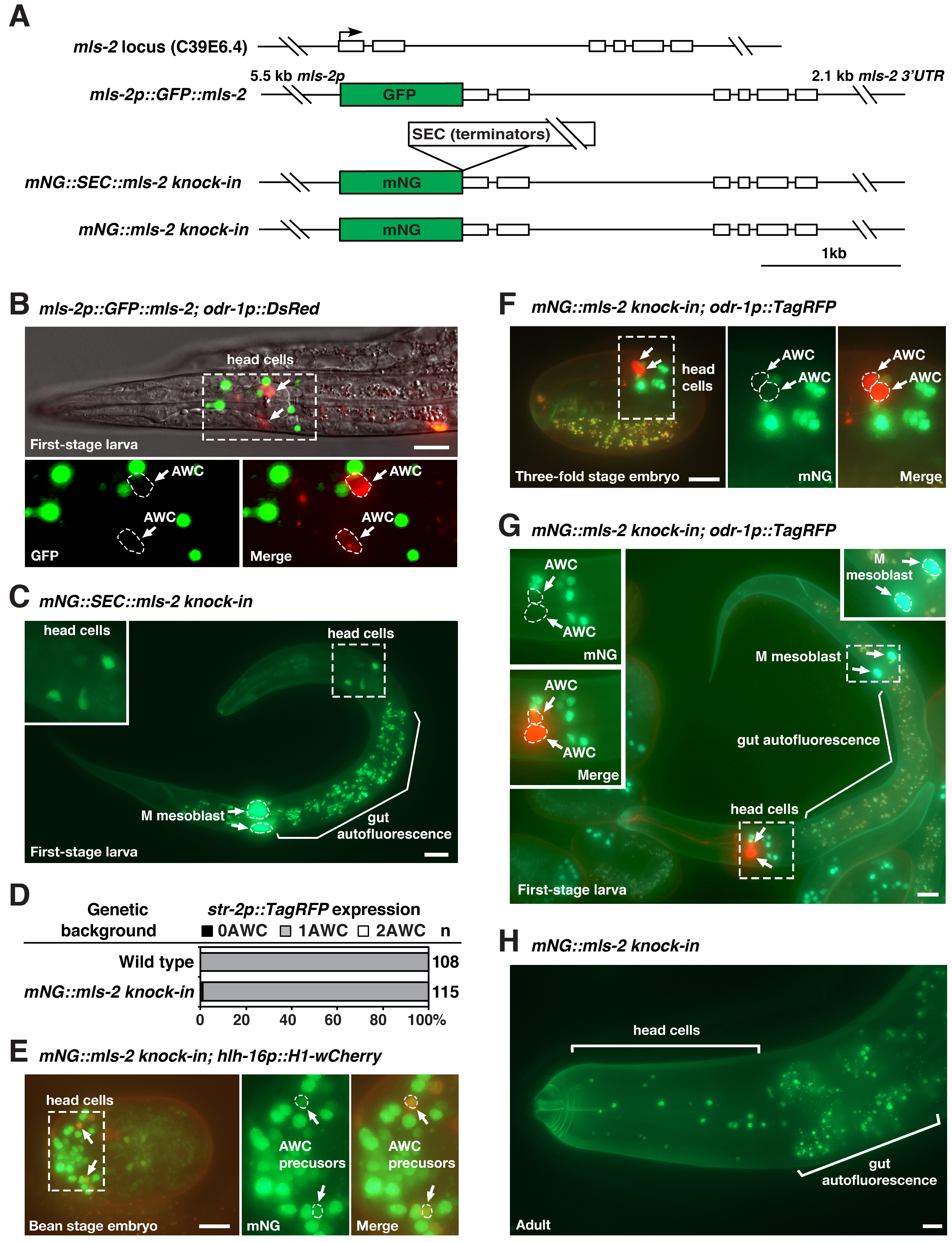
Figure 1. Expression patterns of mls-2p::GFP::mls-2 transgene, mNG::SEC::mls-2 knock-in, and mNG::mls-2 knock-in: (A) Structure of the mls-2 locus, mls-2p::GFP::mls-2 transgene (Jiang et al., 2005), mNG::SEC::mls-2 knock-in, and mNG::mls-2 knock-in. mNG, mNeonGreen. SEC, self-excising cassette containing transcriptional terminators, a dominant roller phenotype marker sqt-1(e1350), Cre driven by a heat shock promoter, and a hygromycin resistance gene. The SEC cassette is flanked by LoxP sites. (B-H) Representative images of GFP::MLS-2 expression from a mls-2p::GFP::mls-2 extrachromosomal transgene in a first-stage larva (B), mNG expression from mNG::SEC::mls-2 knock-in in a first-stage larva (C), and mNG::MLS-2 expression from mNG::mls-2 knock-in animals in different developmental stages (E-H). Integrated transgenes of hlh-16::H1-wCherry and odr-1p::TagRFP (or odr-1p::DsRed) were used as early and late AWC markers, respectively. Insets in panels B, C, E, F, and G are magnified by 2-fold. Scale bar, 10 um. Anterior to the left and ventral to the bottom in lateral or ventrolateral views of the head region in B, C, and F-H; ventral view in E. (D) Expression of the AWCON marker str-2p::TagRFP from an integrated transgene in wild type and mNG::mls-2 knock-in animals. n, total number of animals scored.

Description

The HMX/NKX MLS-2 transcription factor plays a role in the development of the postembryonic mesoderm, CEPsh glia, tube cells of the excretory system, general AWC identity, and AWC asymmetry in C. elegans (Jiang et al., 2005; Yoshimura et al., 2008; Kim et al., 2010; Abdus-Saboor et al., 2012; Hsieh et al., 2021). The expression pattern of MLS-2 protein has been previously examined by immunohistochemical staining with anti-MLS-2 antibodies, transgenes of GFP-tagged MLS-2 (GFP::MLS-2), and mls-2::GFP fosmid reporter lines (Jiang et al., 2005; Yoshimura et al., 2008; Kim et al., 2010; Abdus-Saboor et al., 2012; Walton et al., 2015; Reilly et al., 2020) (Figure 1A). It was shown that GFP::MLS-2 was expressed in the embryonic AWC lineages from automated lineage analysis and was detected transiently in AWC neurons in first-stage larvae (Kim et al., 2010; Abdus-Saboor et al., 2012; Walton et al., 2015). However, we did not detect expression of GFP::MLS-2 transgenes in AWC neurons in late embryos or the first larval stage (Figure 1B).

To determine the expression pattern of endogenous mls-2 locus in AWC, we generated mNG::SEC::mls-2 knock-in and mNG::mls-2 knock-in animals by tagging the 5’ end of endogenous mls-2 coding region with mNG::SEC or mNG using Cas9-triggered homologous recombination (Dickinson et al., 2013; Dickinson et al., 2015; Dickinson and Goldstein, 2016) (Figure 1A). The mNG::SEC::mls-2 knock-in allele is a transcriptional reporter of mls-2, since the self-excising cassette (SEC) contains transcriptional terminators. mNG::SEC::mls-2 knock-in showed diffuse mNG expression in numerous cells in the head and the M mesoblast of first-stage larvae (Figure 1C). The mNG::mls-2 knock-in allele, generated by the SEC excision of mNG::SEC::mls-2 knock-in, is a translational reporter of MLS-2 protein. mNG::mls-2 knock-in animals displayed wild-type AWC asymmetry as determined by the expression of the AWCON marker str-2p::TagRFP (Figure 1D), suggesting that mNG::MLS-2 fusion protein is functional in AWC development. Like GFP::MLS-2 expressed from transgenes, mNG::MLS-2 knock-in was localized in the nucleus of AWC precursor cells in early embryos (Figure 1E) but was not observed in AWC cells in late embryos (Figure 1F) or early-stage larvae (Figure 1G). Our results are consistent with single-cell RNA-seq data showing that mls-2 was briefly expressed at a very low level in AWC during early embryogenesis but not detected in AWC in second-stage larvae (Cao et al., 2017; Packer et al., 2019). It was also shown that MLS-2::GFP expressed from an integrated mls-2::GFP fosmid reporter line was not detected in AWC in late larval stage or young adult-stage using NeuroPAL (Reilly et al., 2020).

Similar to MLS-2 antibody staining and GFP::MLS-2 transgenes (Jiang et al., 2005), mNG::MLS-2 knock-in was localized to the nucleus of a subset of head cells and the M mesoblast in first-stage larvae and adults (Figure 1G and 1H). mNG::SEC::mls-2 knock-in and mNG::mls-2 knock-in strains should help to determine the endogenous expression pattern of mls-2 in different cells during development.

Methods

Request a detailed protocol

mNG::SEC::mls-2 and mNG::mls-2 knock-in were generated using the Cas9-triggered homologous recombination protocol as previously described (Dickinson et al., 2013; Dickinson et al., 2015).

Reagents

Strain Genotype Source
IX1119 oyIs44 [odr-1p::DsRed; lin-15(+)] V; vyEx535 [mls-2p::GFP::mls-2::mls-2 3’UTR (Jiang et al., 2005); ofm-1p::DsRed] This study
IX4507 mls-2(vy247 [mNG::SEC::mls-2 knock-in]) X This study
IX4506 mls-2(vy248 [mNG::mls-2 knock-in]) X This study
RW10588 unc-119(ed3); zuIs178 [his-72(1kb 5′ UTR)::his-72::SRPVAT::GFP::his-72 (1KB 3′ UTR) + 5.7 kb XbaI-HindIII unc-119(+)]; stIs10544 [hlh-16::H1-wCherry::let-858 3′ UTR] Murray et al., 2012
IX5609 stIs10544 [hlh-16::H1-wCherry::let-858 3′ UTR] (Murray et al., 2012); mls-2(vy248 [mNG::mls-2 knock-in]) X This study
IX4894 vyIs56 [odr-1p::TagRFP] III (Cochella et al., 2014); mls-2(vy248 [mNG::mls-2 knock-in]) X This study
IX3212 vyIs68 [str-2p::TagRFP; srsx-3p::GFP] III Cochella et al., 2014

Acknowledgments

We thank Dr. Jun Liu for the plasmid mls-2p::GFP::mls-2::mls-2 3’UTR. The RW10588 strain was provided by the Caenorhadbitis Genetics Center (CGC), which is funded by NIH Office of Research Infrastructure Programs (P40 OD010440).

References

Abdus-Saboor I, Stone CE, Murray JI, Sundaram MV. 2012. The Nkx5/HMX homeodomain protein MLS-2 is required for proper tube cell shape in the C. elegans excretory system. Dev Biol 366: 298-307.
PubMed
Cao J, Packer JS, Ramani V, Cusanovich DA, Huynh C, Daza R, Qiu X, Lee C, Furlan SN, Steemers FJ, Adey A, Waterston RH, Trapnell C, Shendure J. 2017. Comprehensive single-cell transcriptional profiling of a multicellular organism. Science 357: 661-667.
PubMed
Cochella L, Tursun B, Hsieh YW, Galindo S, Johnston RJ, Chuang CF, Hobert O. 2014. Two distinct types of neuronal asymmetries are controlled by the Caenorhabditis elegans zinc finger transcription factor die-1. Genes Dev 28: 34-43.
PubMed
Dickinson DJ, Goldstein B. 2016. CRISPR-Based Methods for Caenorhabditis elegans Genome Engineering. Genetics 202: 885-901.
PubMed
Dickinson DJ, Pani AM, Heppert JK, Higgins CD, Goldstein B. 2015. Streamlined Genome Engineering with a Self-Excising Drug Selection Cassette. Genetics 200: 1035-49.
PubMed
Dickinson DJ, Ward JD, Reiner DJ, Goldstein B. 2013. Engineering the Caenorhabditis elegans genome using Cas9-triggered homologous recombination. Nat Methods 10: 1028-34.
PubMed
Hsieh YW, Xiong R, Chuang CF. 2021. Synergistic roles of homeodomain proteins UNC-62 homothorax and MLS-2 HMX/NKX in the specification of olfactory neurons in Caenorhabditis elegans. Genetics 219: iyab133.
PubMed
Jiang Y, Horner V, Liu J. 2005. The HMX homeodomain protein MLS-2 regulates cleavage orientation, cell proliferation and cell fate specification in the C. elegans postembryonic mesoderm. Development 132: 4119-30.
PubMed
Kim K, Kim R, Sengupta P. 2010. The HMX/NKX homeodomain protein MLS-2 specifies the identity of the AWC sensory neuron type via regulation of the ceh-36 Otx gene in C. elegans. Development 137: 963-74.
PubMed
Murray JI, Boyle TJ, Preston E, Vafeados D, Mericle B, Weisdepp P, Zhao Z, Bao Z, Boeck M, Waterston RH. 2012. Multidimensional regulation of gene expression in the C. elegans embryo. Genome Res 22: 1282-94.
PubMed
Packer JS, Zhu Q, Huynh C, Sivaramakrishnan P, Preston E, Dueck H, Stefanik D, Tan K, Trapnell C, Kim J, Waterston RH, Murray JI. 2019. A lineage-resolved molecular atlas of C. elegans embryogenesis at single-cell resolution. Science 365: eaax1971.
PubMed
Reilly MB, Cros C, Varol E, Yemini E, Hobert O. 2020. Unique homeobox codes delineate all the neuron classes of C. elegans. Nature 584: 595-601.
PubMed
Walton T, Preston E, Nair G, Zacharias AL, Raj A, Murray JI. 2015. The Bicoid class homeodomain factors ceh-36/OTX and unc-30/PITX cooperate in C. elegans embryonic progenitor cells to regulate robust development. PLoS Genet 11: e1005003.
PubMed
Yoshimura S, Murray JI, Lu Y, Waterston RH, Shaham S. 2008. mls-2 and vab-3 Control glia development, hlh-17/Olig expression and glia-dependent neurite extension in C. elegans. Development 135: 2263-75.
PubMed

Funding

This work was funded by a grant, R01GM098026 awarded to CFC, from the National Institute of General Medical Sciences of the National Institutes of Health.

Author Contributions

Rui Xiong: Conceptualization, Methodology, Investigation, Formal analysis, Writing - review and editing
Yi-Wen Hsieh: Conceptualization, Methodology, Investigation, Formal analysis, Writing - review and editing
Chiou-Fen Chuang: Conceptualization, Writing - original draft, Writing - review and editing, Funding acquisition, Supervision.

Reviewed By

Paschalis Kratsios and Jayson Smith

History

Received: January 14, 2022
Revision received: February 9, 2022
Accepted: February 10, 2022
Published: February 22, 2022

Copyright

© 2022 by the authors. This is an open-access article distributed under the terms of the Creative Commons Attribution 4.0 International (CC BY 4.0) License, which permits unrestricted use, distribution, and reproduction in any medium, provided the original author and source are credited.

Citation

Xiong, R; Hsieh, YW; Chuang, CF (2022). mNG-tagged mls-2 knock-in alleles in C. elegans. microPublication Biology. 10.17912/micropub.biology.000529.
Download: RIS BibTeX
microPublication Biology is published by
1200 E. California Blvd. MC 1-43 Pasadena, CA 91125
The microPublication project is supported by
The National Institute of Health -- Grant #: 1U01LM012672-01
microPublication Biology:ISSN: 2578-9430