
Description
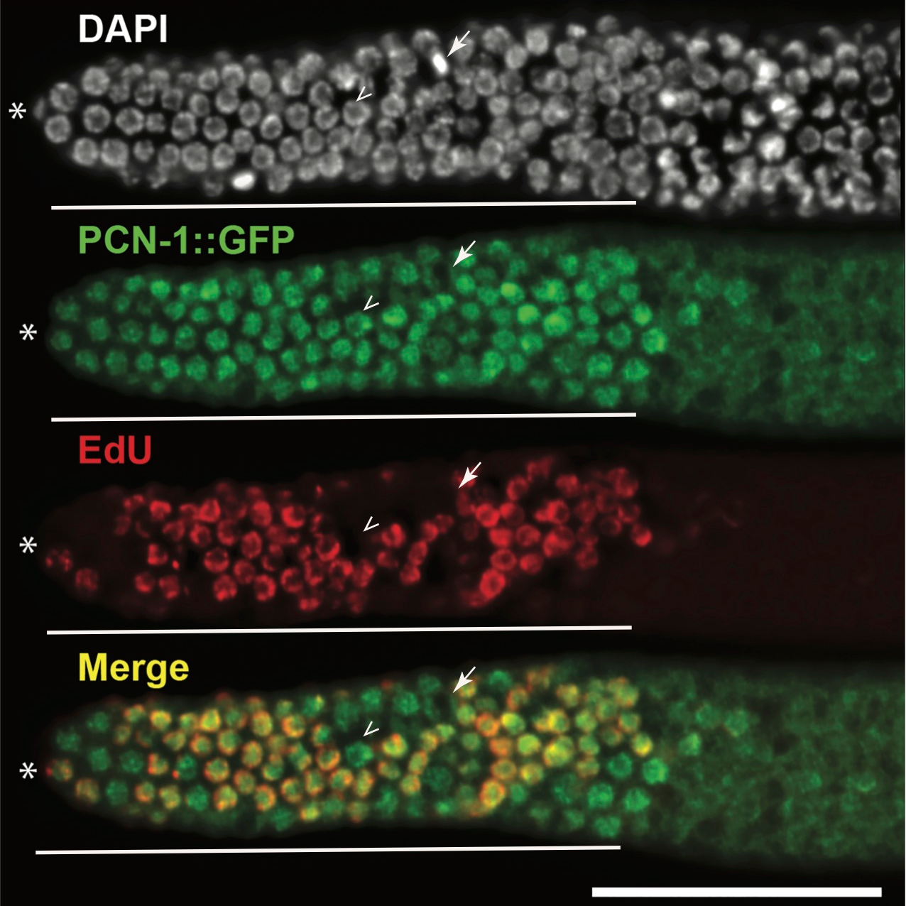
PCNA (proliferating cell nuclear antigen) is the DNA polymerase processivity factor that loads onto the chromatin during S phase of the cell-cycle (Brauchle et al. 2003). Thus, nuclear localization of PCNA (PCN-1 in C. elegans) is used as a marker for the S phase of the cell cycle (Brauchle et al. 2003). GFP::PCN-1 has been shown to label S phase in C. elegans embryo when driven through the germline and embryonic promoter pie-1 (Brauchle et al. 2003). We assayed GFP::PCN-1 (allele isIs17, GZ264 (Brauchle et al. 2003)) as a marker for S phase in adult germline progenitor zone cells. If this reagent were a faithful marker of S phase in germline progenitor zone cells, we would expect nuclear localization during S phase, and nuclear exclusion in the other phases of the cell-cycle, as is the case in the C. elegans embryo. We would also expect a perfect overlap with EdU which marks S phase of the cell cycle. EdU is incorporated in ~55-60% of the adult hermaphroditic wild-type progenitor zone cells (Fox et al. 2011; Furuta et al. 2018). We found that GFP::PCN-1 was nuclear in almost all of the progenitor zone cells, irrespective of whether they were EdU positive or EdU negative (arrowhead, Figure 1). The only cells that excluded GFP::PCN-1 from the nucleus were the metaphase cells (arrow) during M phase, when the nuclear envelope breaks down. Thus, GFP::PCN-1 does not overlap with EdU labeling in adult progenitor zone cells. These data suggest that nuclear localization of GFP::PCN-1 is not a good marker of S phase dynamics in the C. elegans adult germline progenitor cells. This could either be because GFP signal perdures in the nucleus in this context, or that GFP::PCN-1 is nuclear localized throughout the cell cycle in germline progenitor zone cells, unlike in the C. elegans embryo.
Reagents
GFP::PCN-1 worms were grown on nematode growth medium (NGM) plates with E. coli OP50 bacteria. Adult 24 hours past mid-L4 hermaphroditic worms were incubated with 200 µM of EdU solution for 10 minutes at room temperature in the dark. After the EdU soaking, the animals were dissected and extruded germlines processed using the Click-iT® Plus EdU Alexa Fluor® 594 Imaging Kit (ThermoFisher Scientific, catalog number C10639) per the manufacturer’s recommendations (Furuta et al. 2018). After EdU processing, the germlines were treated with anti-GFP antibody (developed in the Arur Lab), to visualize GFP::PCN-1, followed by treatment with donkey-anti-Rabbit-488 secondary antibody (Thermofisher Scientific, catalog number A21260). Antibody treated germlines were stained with 2μg/ml DAPI (4′,6-diamidine-2-phenylindole dihydrochloride) and finally suspended in 10μl of Vectashield (antifade agent).
References
Funding
Work in the Arur Lab is funded through NIH GM98200, CPRIT RP160023, ACS DDC-RSG-14-044-01 and Anna Fuller Foundation. SA is a fellow of the Andrew Sabin Family Foundation at University of Texas MD Anderson Cancer Center.
Reviewed By
Ariz MohammadHistory
Received: May 23, 2018Accepted: May 29, 2018
Published: May 30, 2018
Copyright
© 2018 by the authors. This is an open-access article distributed under the terms of the Creative Commons Attribution 4.0 International (CC BY 4.0) License, which permits unrestricted use, distribution, and reproduction in any medium, provided the original author and source are credited.Citation
Uruta, T; Arur, S (2018). GFP::PCN-1 does not reliably mark S phase in C. elegans adult germline progenitor zone cells. microPublication Biology. 10.17912/W20W9G.Download: RIS BibTeX
