microPublication

Get Your Data Out, Be Cited

  • About
    • Editorial Policies
      • Editorial Staff
      • Editorial Board
      • Criteria For Publication
      • Publishing Information
      • Data Sharing Policy
    • For Authors
      • Preparation And Submission Of A Manuscript
      • Peer Review Process
      • Following Acceptance
      • Appeals
    • For Reviewers
    • Why micropublish?
  • Submit a microPublication
  • Journals
    • microPublication Biology
      • Editorial Board
  • microPublications
    • Biology
      • Species
        • Arabidopsis
        • C. elegans
        • D. discoideum
        • Drosophila
        • Human
        • Mouse
        • S. cerevisiae
        • S. pombe
        • Xenopus
        • Zebrafish
      • Categories
        • Phenotype Data
        • Methods
        • Expression Data
        • Genotype Data
        • Integrations
        • Genetic Screens
        • Models of Human Disease
        • Software
        • Interaction data
        • Database Updates
        • Electrophysiology Data
        • Phylogenetic Data
        • Science and Society
        • Biochemistry
  • Contact
  • More
    • Archives
    • FAQs
    • Newsletter
microPublication / Biology / The C. elegans CEPsh glia...
The C. elegans CEPsh glia are largely dispensable for stress-induced sleep
Rony Soto1 and Cheryl Van Buskirk1
1Department of Biology, California State University Northridge, Northridge CA USA 91330
Correspondence to: Cheryl Van Buskirk (cheryl.vanbuskirk@csun.edu)
Figure 1: CEPsh ablation mildly impairs UV–SIS, but heat–SIS and Cry5B–SIS are intact. (A–C) Examination of wild-type N2, ALA–defective ceh-17(np1), and CEPsh glia–ablated (GA) lines during stress-induced sleep (SIS). Pre-fertile adult animals were exposed to conditions known to trigger SIS and examined for behavioral quiescence as described in Methods. Whereas reduction of ALA neuron function impairs all forms of SIS, ablation of the CEPsh glia has no significant effect on heat–SIS (A) nor Cry5B toxin–induced sleep (B). Two of the three glia–ablated lines show mildly reduced SIS following UVB light exposure (C). (A) ns = not significant, *p<0.05 vs. wild type, one-way ANOVA of area under the curve with Dunnett’s multiple comparisons test; mean and SEM of three trials of 25 animals each are shown. (B,C) ns = not significant, *p<0.05, **p<0.01,***p<0.001 vs. wild type, one-way ANOVA with Dunnett's multiple comparisons test; each data point represents the fraction of animals quiescent in one trial (n=25), and the mean of three trials is indicated.

Description

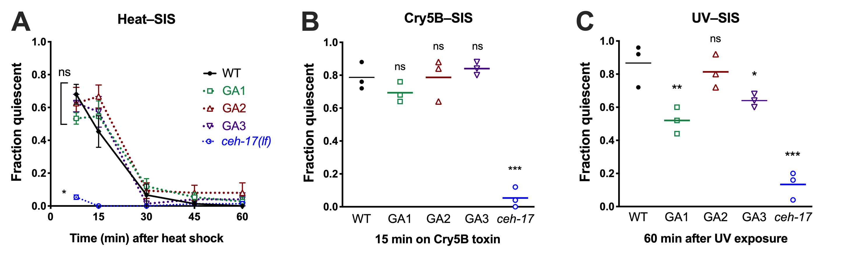
Across species, sleep is increased following exposure to damaging conditions, a phenomenon known as stress-induced sleep (SIS) (Hill et al. 2014; Lenz et al. 2015; Zada et al. 2019). In C. elegans, SIS is dependent on the ALA interneuron (Hill et al. 2014; Nelson et al. 2014), which promotes a coordinated quiescent state through the collective action of several neuropeptides (Nelson et al. 2014; Nath et al. 2016). Recently it has been shown that ALA ablation can suppress phenotypes associated with loss of the cephalic sensilla sheath (CEPsh) glia, such as prolonged larval development and locomotor pausing in adults (Katz et al. 2018), indicating that these glia attenuate certain aspects of ALA function. The CEPsh glia form a tubular structure surrounding the sensory endings of the CEP neurons and also extend thin processes that sheath the nerve ring, including the synapse between ALA and the postsynaptic AVE, a major locomotor interneuron. While the ALA is likely to promote SIS via peptidergic rather than synaptic signaling (Nelson et al. 2014; Nath et al. 2016), we wished to determine whether CEPsh glia ablation may impact the SIS-promoting function of ALA. To this end, we examined three independent lines that genetically ablate the four CEPsh glial cells (Katz et al. 2018), and compared the SIS responses of these strains to wild type and to ALA specification-defective ceh-17(np1) mutant animals. We examined three conditions known to trigger ALA-dependent sleep: noxious heat, pore-forming Cry5B toxin, and ultraviolet light exposure (Figure 1). We found that while the glia-ablated (GA) lines displayed a trend toward enhanced heat-SIS at the 15min time point, there were no significant differences in heat-SIS between the GA lines and wild-type. The GA lines also showed wild-type Cry5B-SIS. Surprisingly, two of the GA lines showed reduced UV-SIS, rather than the enhanced sleep that would be predicted if the CEPsh glia attenuate ALA’s peptidergic function. Our data indicate that CEPsh glia are largely dispensable for stress-induced sleep, but may modulate the UV–SIS response. As UV exposure elicits a delayed sleep response relative to other SIS triggers, we speculate that UV damage may activate ALA in a manner that is distinct, and partially supported by glial function.

Methods

Request a detailed protocol

CEPsh glia were ablated at the L1 stage by expression of a reconstituted caspase-3 gene from the hlh-17 promoter (Katz et al. 2018). Behavior was assessed in well-fed (on E. coli OP50) pre-fertile adult animals on NGM agar plates. Quiescence was defined as complete immobility and lack of pharyngeal pumping during a 5 second observation under a stereomicroscope at 375x magnification with experimenter blind to genotype. We observed that immobility was accompanied by feeding quiescence in all cases, but not vice versa, i.e., ‘fraction quiescent’ is equivalent to ‘fraction immobile’. For heat-SIS, 12 ml agar plates were sealed with parafilm and placed upright in a 37°C water bath for 11 minutes, then placed on ice for 2 minutes to return them to room temperature (Goetting et al. 2018). For Cry5B-SIS, animals were placed onto NGM plates containing Cry5B-expressing bacteria (Hill et al. 2014) and examined for SIS 15 minutes later in the presence of Cry5B. For UV-SIS, plates were placed lid-side down on a 302 nm 60 mW ultraviolet (UVB) light source for 50 seconds and examined 60 minutes later for SIS, as UV-induced sleep takes longer to set in than other forms of SIS (DeBardeleben et al. 2017; Goetting et al. 2018).

Reagents

Wild-type N2 and IB16 ceh-17(np1) from the CGC; OS3537 (GA1) nsIs168 (Phlh-17::recCaspase-3, Punc-122::GFP, Pptr-10::myrRFP), OS3540 (GA2) nsIs171 (Phlh-17::recCaspase-3, Punc-122::GFP), and OS3549 (GA3) nsIs180 (Phlh-17::recCaspase-3, Punc-122::GFP) from M. Katz; Cry5B toxin from Raffi Aroian. All reagents are available from our lab.

Acknowledgments

Many thanks to Menachem Katz for strains and for helpful comments on the manuscript.

References

DeBardeleben, H.K., Lopes, L.E., Nessel, M.P., and Raizen, D.M. (2017). Stress-induced sleep after exposure to ultraviolet light is promoted by p53 in Caenorhabditis elegans. Genetics 207, 571–582.
https://doi.org/10.1534/genetics.117.300070
Hill, A.J., Mansfield, R., Lopez, J.M.N.G., Raizen, D.M., and Van Buskirk, C. (2014). Cellular Stress Induces a Protective Sleep-like State in C. elegans. Curr. Biol. 24, 2399–2405.
https://doi.org/10.1016/j.cub.2014.08.040
Goetting, D. L., Soto, R., and Van Buskirk, C. (2018). Food-Dependent Plasticity in Caenorhabditis elegans Stress-Induced Sleep Is Mediated by TOR-FOXA and TGF-beta Signaling. Genetics 209, 1183–1195.
https://doi.org/10.1534/genetics.118.301204
Katz, M., Corson, F., Iwanir, S., and Shaham, S. (2018). Glia Modulate a Neuronal Circuit for Locomotion Suppression during Sleep in C. elegans. Cell Rep. 22, 2575-2583.
https://doi.org/10.1016/j.celrep.2018.02.036
Lenz, O., Xiong, J., Nelson, M. D., Raizen, D. M., and Williams, J. A. (2015). FMRFamide signaling promotes stress-induced sleep in Drosophila. Brain, Behav. Immun. 47, 141–148.
https://doi.org/10.1016/j.bbi.2014.12.028
Nath, R. D., Chow, E. S., Wang, H., Schwarz, E. M., and Sternberg, P. W. (2016). C. elegans Stress-Induced Sleep Emerges from the Collective Action of Multiple Neuropeptides. Curr. Biol. 26, 2446–2455.
https://doi.org/10.1016/j.cub.2016.07.048
Nelson, M. D., Lee, K. H., Churgin, M. A., Hill, A. J., Van Buskirk, C., Fang-Yen, C., and Raizen, D. M. (2014). FMRFamide-like FLP-13 Neuropeptides Promote Quiescence following Heat Stress in Caenorhabditis elegans. Curr. Biol. 24, 2406–2410.
https://doi.org/10.1016/j.cub.2014.08.037
Zada, D., Bronshtein, I., Lerer-Goldshtein, T., Garini, Y., and Appelbaum, L. (2019). Sleep increases chromosome dynamics to enable reduction of accumulating DNA damage in single neurons. Nat. Commun. 10, 895.
https://doi.org/10.1038/s41467-019-08806-w

Funding

C.V.B. is supported by the National Science Foundation Faculty Early Career Development Program (IOS#1553673). The Caenorhabditis Genetics Center (CGC) is funded by the National Institutes of Health Office of Research Infrastructure Programs (P40 OD-010440).

Author Contributions

Rony Soto: Conceptualization, Formal analysis, Investigation, Writing - review and editing
Cheryl Van Buskirk: Funding acquisition, Supervision, Writing - original draft, Writing - review and editing.

Reviewed By

David Raizen

History

Received: May 6, 2020
Revision received: May 28, 2020
Accepted: May 30, 2020
Published: May 30, 2020

Copyright

© 2020 by the authors. This is an open-access article distributed under the terms of the Creative Commons Attribution 4.0 International (CC BY 4.0) License, which permits unrestricted use, distribution, and reproduction in any medium, provided the original author and source are credited.

Citation

Soto, R; Van Buskirk, C (2020). The C. elegans CEPsh glia are largely dispensable for stress-induced sleep. microPublication Biology. 10.17912/micropub.biology.000261.
Download: RIS BibTeX
microPublication Biology is published by
1200 E. California Blvd. MC 1-43 Pasadena, CA 91125
The microPublication project is supported by
The National Institute of Health -- Grant #: 1U01LM012672-01
microPublication Biology:ISSN: 2578-9430